. 2011 Jun;44(6):755-60.
doi: 10.1165/rcmb.2010-0020oc.

Mucin production during prenatal and postnatal murine lung development

Affiliations

Mucin production during prenatal and postnatal murine lung development

Michelle G Roy et al. Am J Respir Cell Mol Biol. 2011 Jun.

Abstract

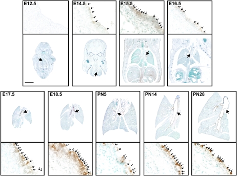
Mucus is a protective gel that lines respiratory tract surfaces. To identify potential roles for secreted gel--forming mucins in lung development, we isolated murine lungs on embryonic days (E) 12.5-18.5, and postnatal days (PN) days 5, 14, and 28. We measured the mucin gene expression by quantitative RT-PCR, and localization by histochemical and immunohistochemical labeling. Alcian blue/periodic acid--Schiff--positive cells are present from E15.5 through PN28. Muc5b transcripts were abundant at all time points from E14.5 to PN28. By contrast, transcript levels of Muc5ac and Muc2 were approximately 300 and 85,000 times lower, respectively. These data are supported by immunohistochemical studies demonstrating the production and localization of Muc5ac and Muc5b protein. This study indicates that mucin production is prominent in developing murine lungs and that Muc5b is an early, abundant, and persistent marker of bronchial airway secretory cells, thereby implicating it as an intrinsic component of homeostatic mucosal defense in the lungs.


Figures

Figure 1.
Figure 1.
Alcian blue/periodic acid–Schiff (AB-PAS)–positive cells are prominent in prenatal and postnatal developing murine lungs. Tissue sections (5 μm) were removed from whole embryos (E) and isolated lungs of postnatal (PN) wild-type C57/BL6 mice isolated at the indicated time points, and stained with AB-PAS. Large arrows in low-magnification images (top) identify airway sites shown in high magnification (below). Small arrows (below) identify positively stained cells. Scale bar, 2 mm for low magnification, and 30 μm for high magnification.
Figure 2.
Figure 2.
Quantitative RT-PCR analysis demonstrates the expression of Muc5ac and Muc5b in the developing murine lung. Gene-specific probes were used to measure concentrations of the mucins Muc2 (solid circles), Muc5ac (solid squares), and Muc5b (solid triangles), and of Clara cell secretory protein (Scgb1a1) (open circles), compared with γ-actin. Data represent means ± standard errors (n = 3–8 animals/time point). Data were analyzed by ANOVA. P < 0.05 was considered significant. **Identifies significance between concentrations of Muc5b mRNA and those of Muc5ac and Muc2. Identifies significance between Muc5ac and Muc2. Identifies significance between embryonic day (E) 14.5 and later values. Differences in concentrations of Muc5ac and Muc2 were not significant over time. PN, postnatal day.
Figure 3.
Figure 3.
Muc5b protein is constitutively produced in prenatal and postnatal developing murine lungs. Immunostaining for Muc5b was performed using polyclonal rabbit anti-mouse Muc5b antibody (1:10,000), and detected with a peroxidase-conjugated goat anti-rabbit antibody (1:200) and 3,3′-diaminobenzidine (DAB). Large arrows in low-magnification images (top) identify airway sites shown in high magnification (below). Small arrows (below) identify positively stained cells. Scale bar, 2 mm for low magnification, and 30 μm for high magnification.
Figure 4.
Figure 4.
Muc5ac protein is briefly produced in postnatal developing murine lungs. Immunostaining for Muc5ac was performed using polyclonal chicken anti-mouse Muc5ac peptide chicken polyclonal antibody (1:2,000), and detected with a biotinylated goat anti-chicken antibody (1:500) and DAB. Large arrows in low-magnification images (top) identify airway sites shown in high magnification (below). Small arrows (below) identify positively stained cells. Scale bar, 2 mm for low magnification, and 30 μm for high magnification.

References

    1. Warburton D, Schwarz M, Tefft D, Flores-Delgado G, Anderson KD, Cardoso WV. The molecular basis of lung morphogenesis. Mech Dev 2000;92:55–81. - PubMed
    1. Evans CM, Koo JS. Airway mucus: the good, the bad, the sticky. Pharmacol Ther 2009;121:332–348. - PMC - PubMed
    1. Rose MC, Voynow JA. Respiratory tract mucin genes and mucin glycoproteins in health and disease. Physiol Rev 2006;86:245–278. - PubMed
    1. Thornton DJ, Rousseau K, McGuckin MA. Structure and function of the polymeric mucins in airways mucus. Annu Rev Physiol 2008;70:459–486. - PubMed
    1. Whitsett JA, Weaver TE. Hydrophobic surfactant proteins in lung function and disease. N Engl J Med 2002;347:2141–2148. - PubMed

Publication types