. 2011 Jun 7;30(14):2948-61.
doi: 10.1038/emboj.2011.185.

An infrequent molecular ruler controls flagellar hook length in Salmonella enterica

Affiliations

An infrequent molecular ruler controls flagellar hook length in Salmonella enterica

Marc Erhardt et al. EMBO J. .

Abstract

The bacterial flagellum consists of a long external filament connected to a membrane-embedded basal body at the cell surface by a short curved structure called the hook. In Salmonella enterica, the hook extends 55 nm from the cell surface. FliK, a secreted molecular ruler, controls hook length. Upon hook completion, FliK induces a secretion-specificity switch to filament-type substrate secretion. Here, we demonstrate that an infrequent ruler mechanism determines hook length. FliK is intermittently secreted during hook polymerization. The probability of the specificity switch is an increasing function of hook length. By uncoupling hook polymerization from FliK expression, we illustrate that FliK secretion immediately triggers the specificity switch in hooks greater than the physiological length. The experimental data display excellent agreement with a mathematical model of the infrequent ruler hypothesis. Merodiploid bacteria expressing simultaneously short and long ruler variants displayed hook-length control by the short ruler, further supporting the infrequent ruler model. Finally, the velocity of FliK secretion determines the probability of a productive FliK interaction with the secretion apparatus to change secretion substrate specificity.


Conflict of interest statement

The authors declare that they have no conflict of interest.

Figures

Figure 1
Figure 1
(A) Schematic of axial components of the bacterial flagellum. The structure of the bacterial flagellum can be divided into three parts: (i) the basal-body structure that harbours the flagellar-specific type III secretion apparatus at the base; (ii) the hook that functions as a flexible coupling structure between the basal body and (iii) the rigid filament. Stator elements (Mot proteins) that span the inner membrane and apply torque to the C-ring in response to transmembrane proton flow are not shown. Rod-hook length is determined by a molecular ruler, FliK, that in turn induces a switch in secretion specificity from rod-hook-type to filament-type substrates upon hook completion, presumably by interaction with FlhB, a component of the type III secretion apparatus at the base of the structure. (B) Schematic of experimental outline. An overnight culture of a strain expressing the flagellar master operon flhDC from a Tc-inducible PtetA promoter is diluted into fresh LB and grown for 3 h. After 3 h growth, flagellar gene expression is induced by addition of Tc and 30 min after induction transcription of Class 3 promoters is observed, which indicates HBB completion (Karlinsey et al, 2000). Here, we uncoupled FliK expression from flagellar genes expression to analyse the effects of late FliK induction on switching from HBB-type secretion to filament-type secretion in a strain deleted for its native fliK gene and expressing fliK from the inducible ParaBAD promoter (PtetA-flhD+C+ ParaBAD-fliK+ ΔfliK). In the first sample (‘wild-type’), flagellar gene expression and fliK expression are induced simultaneously by addition of Tc to induce the flagellar master regulator flhDC (PtetA-flhD+C+) and Ara to induce fliK expression (ParaBAD-fliK+) resulting in hooks of wild-type length. In the second sample (‘polyhook’), only Tc is added to induce flagellar gene expression, giving rise to polyhooks because FliK is not induced. In the third sample (‘late fliK induction’), flagellar gene expression is induced for 45 min without FliK expression. This allows for hook-length growth beyond the physiological length. Afterwards, fliK is induced by addition of Ara and the culture grown for an additional 30 min to allow for induction of the secretion-specificity switch and filament assembly.
Figure 2
Figure 2
FliK induces secretion-specificity switch in hooks >wt length. (A) Immunostaining of assembled HBB complexes and filaments. ‘WT’: simultaneous induction of fliK and flagellar genes expression; ‘polyhook’: flagellar genes were induced without induction of fliK; ‘late FliK’: late fliK induction after 45 min of flagellar genes expression for 10 and 30 min, respectively. Tc was not removed prior to addition of Ara. Representative fluorescent microscopy images of strain TH16941 (PtetA-flhD+C+ ParaBAD-fliK+ ΔfliK flgE∷3xHA) are shown. DNA (blue), hooks (white) and filaments (green). Scale bar=2 μm. (B) Time lapse of fliC transcription after fliK induction. FliK expression in strain TH17502 harbouring a fliC-lac reporter was induced late after 45 min of flagellar genes expression. β-galactosidase activity was assayed according to Materials and methods. The inducer of flagellar gene expression, Tc, was not removed prior to addition of Ara. Data are presented as mean±s.d. of three independent, biological replicates. (C) Filament-length distribution after 10 and 30 min of late fliK induction compared with the simultaneous induction of fliK and flagellar genes expression (‘WT’). Number of filaments measured: ‘WT’=34, ‘10 min’=36 and ‘30 min’=47.
Figure 3
Figure 3
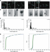
Late FliK secretion induces secretion-specificity switch in elongated hooks. Left panels (WT): simultaneous induction of fliK and flagellar genes expression. Middle panels (polyhook): flagellar genes were induced without induction of fliK. Right panels (late FliK): late fliK induction after 45 min of flagellar genes expression. (A) Representative fluorescent microscopy images of strain TH16941 (PtetA-flhD+C+ ParaBAD-fliK+ ΔfliK flgE∷3xHA). Tc was removed prior to addition of Ara to prevent formation of nascent HBBs. Number of cells counted for the presence/absence of HBB–filament complexes: ‘WT’=400, ‘polyhook’=642, ‘late FliK’=321. Fraction of HBBs with attached filaments is given in the upper left corner. DNA (blue), hooks (white) and filaments (green). Scale bar=2 μm. (B) Representative electron micrograph images of hooks isolated from strain TH16791 (PtetA-flhD+C+ ParaBAD-fliK+ ΔfliK). Scale bar=50 nm. (C) Histogram of measured hooks of strain TH16791. Number of measured hooks: ‘WT’=66, ‘polyhook’=229, ‘late FliK’=228. (D) CDF of hooks measured for TH16791. Measured hook lengths shown as asterisks and Pi(L) (solid curve) computed from equation 8 using L*=470 nm.
Figure 4
Figure 4
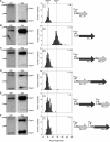
Late induction of fliK at varying times T0. Strain TH16791 (PtetA-flhD+C+ ParaBAD-fliK+ ΔfliK) was grown in the presence of Tc (inducer of flagellar genes) for (A) 45 min, (B) 55 min and (C) 65 min. Afterwards, fliK expression was induced by addition of Ara for a total sample time of 80 min. Left panels: histogram of measured hooks. Number of measured hooks: ‘T0=45 min’=286, ‘T0=55 min’=461, ‘T0=65 min’=134. Right panels: CDF of measured hooks, data shown as asterisks and Pi(L) (solid curve) computed from equation 8 using L*=600 nm (T0=45 min), 440 nm (T0=55 min) and 260 nm (T0=65 min).
Figure 5
Figure 5
Late secretion of short FliK363 and long FliK570. FliK363 (left panels) and FliK570 (right panels) expression was induced late after 45 min of flagellar genes expression. (A) Representative fluorescent microscopy images of strain TH17012 (PtetA-flhD+C+ ParaBAD-fliKΔ161–202 ΔfliK flgE∷3xHA) and TH17011 (PtetA-flhD+C+ ParaBAD-fliK(140′-yscP(217–381)-141fliK) ΔfliK flgE∷3xHA). Tc was removed prior to addition of Ara to prevent formation of nascent HBBs. Number of cells counted for the presence/absence of HBB–filament complexes: ‘late FliK363’=367, ‘late FliK570’=180. Fraction of HBBs with attached filaments is given in the upper left corner. DNA (blue), hooks (white) and filaments (green). Scale bar=2 μm. (B) Representative electron micrograph images of hooks isolated from strain TH16997 (PtetA-flhD+C+ ParaBAD-fliKΔ161–202 ΔfliK) and TH16996 (PtetA-flhD+C+ ParaBAD-fliK(140′-yscP(217–381)-′141fliK) ΔfliK). Scale bar=50 nm. (C) Histogram of measured hooks of strain TH16997 (FliK363) and TH16996 (FliK570). Number of measured hooks: ‘late FliK363’=80, ‘late FliK570’=244. (D) CDF of hooks measured for strain TH16997 and TH16996. Measured hook lengths shown as asterisks and Pi(L) (solid curve) computed from equation 8 using L*=440 nm (FliK363) and L*=540 nm (FliK570).
Figure 6
Figure 6
Velocity of FliK secretion is dependent on hook length. Cellular FliM (left panels), cellular FliK (middle panels) and extracellular FliK levels (right panels). Expression of flagellar genes and fliK was induced as outlined in Materials and methods. Residual Tc was washed out before induction of FliK and samples were taken at the time points indicated. Representative western blots are shown. (A) Top: FliK secretion in strain that produces elongated hooks (strain TH16791). Bottom: FliK secretion in the absence of the hook (strain TH17069). (B) Top: FliK secretion in a long hook strain that is unable to switch to late-substrate secretion mode (TH17076, FlhB autocleaveage defective mutant). Bottom: FliK secretion in the switch, hook strain TH17112. (C) Relative levels of secreted FliK normalized against intracellular FliM of TH16791 and TH17069 from the blot shown in (A). (D) Relative levels of secreted FliK normalized against intracellular FliM of TH17112 and TH17076 from the blot shown in (B).
Figure 7
Figure 7
Hook-length distribution of merodiploid strains co-expressing short and long FliK variants. Left panels: pellet and supernatant fractions were analysed by immunoblotting using polyclonal anti-FliK antibodies. Middle panels: hook-length distribution of purified HBB complexes of the different strains used. The average hook length±s.d. is given in the upper left corner and was obtained by non-linear regression analysis of the Gaussian distribution. The average hook lengths for FliK363(art) (nucleotide sequence given in Supplementary Figure S5) and FliK570 are indicated by the dashed line. Number of measured hooks for each construct is indicated in the figure. Right panels: schematic of the various genetic constructs. (A) Strain TH7556 expressing the artificial FliK363(art) variant from ParaBAD. (B) Strain TH16996 expressing FliK570 from ParaBAD. (C) Strain TH17555 expressing FliK570–FliK363(art) from ParaBAD. (D) Strain TH17557 expressing FliK363(art)–FliK570 from ParaBAD. (E) Strain TH17457 expressing FliK570 from ParaBAD and FliK363 from PfliF. (F) Strain TH17458 expressing FliK363 from ParaBAD and FliK570 from PfliF.
Figure 8
Figure 8
Model of hook-length determination by the infrequent ruler mechanism. (A) FliK is intermittently secreted during hook polymerization. During FliK secretion, hook polymerization temporarily halts and the N-terminus of FliK interacts with assembled FlgE and FlgD subunits during its secretion. The lack of interactions in short hooks or the folding of the secreted FliK N-terminus as it exits the secretion channel rapidly pulls the FliK molecule past FlhB through the channel without induction of the secretion-specificity switch (see text for details). (B) FliK is secreted outside of the cell and hook polymerization continues. (C) The hook has grown to the physiological (or longer) length (here: 55 nm). A new FliK molecule is secreted and this time secretion is slower because of more frequent interactions of the FliK N-terminus with assembled hook subunits. Additionally, the N-terminus of FliK is not yet secreted outside of the cell, where its fold might enhance secretion. (D) The C-terminus of FliK is now closely aligned to FlhB and the slower rate of FliK secretion allows for sufficient time for a productive FliK–FlhB interaction that induces the secretion-specificity switch (indicated by a yellow star in the figure).

References

    1. Agrain C, Sorg I, Paroz C, Cornelis GR (2005) Secretion of YscP from Yersinia enterocolitica is essential to control the length of the injectisome needle but not to change the type III secretion substrate specificity. Mol Microbiol 57: 1415–1427 - PubMed
    1. Aizawa SI, Dean GE, Jones CJ, Macnab RM, Yamaguchi S (1985) Purification and characterization of the flagellar hook-basal body complex of Salmonella typhimurium. J Bacteriol 161: 836–849 - PMC - PubMed
    1. Berg HC, Anderson RA (1973) Bacteria swim by rotating their flagellar filaments. Nature 245: 380–382 - PubMed
    1. Blocker A, Komoriya K, Aizawa S (2003) Type III secretion systems and bacterial flagella: insights into their function from structural similarities. Proc Natl Acad Sci USA 100: 3027–3030 - PMC - PubMed
    1. Chevance FFV, Hughes KT (2008) Coordinating assembly of a bacterial macromolecular machine. Nat Rev Microbiol 6: 455–465 - PMC - PubMed

Publication types

Substances