. 2011 Oct;19(10):1074-81.
doi: 10.1038/ejhg.2011.86. Epub 2011 Jun 8.

A dominant mutation in RPE65 identified by whole-exome sequencing causes retinitis pigmentosa with choroidal involvement

Affiliations

A dominant mutation in RPE65 identified by whole-exome sequencing causes retinitis pigmentosa with choroidal involvement

Sara J Bowne et al. Eur J Hum Genet. 2011 Oct.

Erratum in

  • Eur J Hum Genet. 2011 Oct;19(10):1109

Abstract

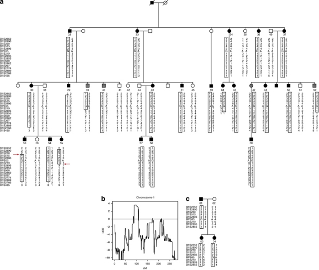
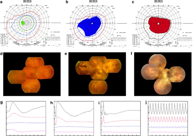
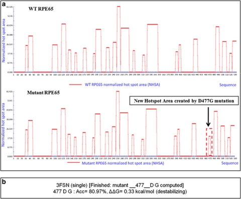
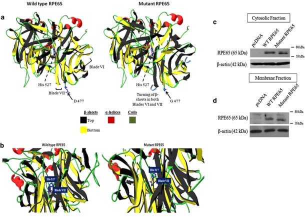
Linkage testing using Affymetrix 6.0 SNP Arrays mapped the disease locus in TCD-G, an Irish family with autosomal dominant retinitis pigmentosa (adRP), to an 8.8 Mb region on 1p31. Of 50 known genes in the region, 11 candidates, including RPE65 and PDE4B, were sequenced using di-deoxy capillary electrophoresis. Simultaneously, a subset of family members was analyzed using Agilent SureSelect All Exome capture, followed by sequencing on an Illumina GAIIx platform. Candidate gene and exome sequencing resulted in the identification of an Asp477Gly mutation in exon 13 of the RPE65 gene tracking with the disease in TCD-G. All coding exons of genes not sequenced to sufficient depth by next generation sequencing were sequenced by di-deoxy sequencing. No other potential disease-causing variants were found to segregate with disease in TCD-G. The Asp477Gly mutation was not present in Irish controls, but was found in a second Irish family provisionally diagnosed with choroideremia, bringing the combined maximum two-point LOD score to 5.3. Mutations in RPE65 are a known cause of recessive Leber congenital amaurosis (LCA) and recessive RP, but no dominant mutations have been reported. Protein modeling suggests that the Asp477Gly mutation may destabilize protein folding, and mutant RPE65 protein migrates marginally faster on SDS-PAGE, compared with wild type. Gene therapy for LCA patients with RPE65 mutations has shown great promise, raising the possibility of related therapies for dominant-acting mutations in this gene.


Figures

Figure 1
Figure 1
Linkage analysis in TCD-G (a) TCD-G pedigree and STR typing data for linkage region on chromosome 1. (b) TCD-G genome wide linkage analysis results for chromosome 1. (c) TCD-H pedigree and STR typing data.
Figure 2
Figure 2
TCD-G clinical evaluation (a) Goldmann perimetry for the right eye from a very mildly affected individual, showing slight loss of superior field to the large IV4e target, (b) a moderately severely affected individual illustrating the complete mid-peripheral ring scotoma to the intermediate sized I4e target, and (c) a severely affected individual illustrating a complete mid-peripheral ring scotoma to the large IV4e target. (d) Composite fundus photographs from the right eye of a mildly affected individual showing bone spicule and nummular intraretinal pigmentary deposits in the mid-periphery. The area of pigment deposition also shows thinning of the underlying retinal pigment epithelium, (e) a moderately severely affected individual showing extensive areas of chorioretinal atrophy with pigment clumping particularly evident temporally, and (f) a severely affected individual. Extensive, diffuse chorioretinal atrophy is evident, affecting in particular the macular area. Nummular intraretinal pigmentary deposits are visible. This fundus picture bears a superficial resemblance to that seen in choroideremia. (g) Rod-isolated electroretinographic responses from a normal individual (black), a mildly affected patient from TCD-G (red), a moderately severely affected patient (blue), and a severely affected patient (purple). Note the reduction in amplitude, but normal timing of the response, from the mildly affected patient compared with the markedly reduced amplitude, and delayed response from the moderately affected person, and the non-recordable response from the severely affected individual. The late-downward deflection in the purple tracing is an artefact due to the patient blinking. The vertical amplitude scale is 250 μV per division. The horizontal timescale is 50 ms per division. (h) Mixed rod and cone electroretinographic responses from a normal individual (black), a mildly affected patient from TCD-G (red), a moderately severely affected patient (blue), and a severely affected patient (purple). Note the reduction in amplitude, but normal timing of the response, from the mildly affected patient compared with the markedly reduced amplitude, and delayed response from the moderately affected person, and the non-recordable response from the severely affected individual. The red and blue responses show relatively better preservation of the negative a-wave compared with the later positive b-wave. The vertical amplitude scale is 250 μV per division. The horizontal timescale is 50 ms per division. (i) 0.5 Hz cone electroretinographic responses from a normal individual (black), a mildly affected patient from TCD-G (red), a moderately affected patient (blue), and a severely affected patient (purple). Note the reduction in amplitude, but normal timing of the response, from the mildly affected patient compared with the markedly reduced amplitude, and delayed response from the moderately affected person, and the non-recordable response from the severely affected individual. The vertical amplitude scale is 100 μV per division. The horizontal timescale is 50 ms per division. (j) 30 Hz cone electroretinographic responses from a normal individual (black), a mildly affected patient from TCD-G (red), a moderately severely affected patient (blue), and a severely affected patient (purple). Note the reduction in amplitude, but normal timing of the response, from the mildly affected patient compared with the markedly reduced amplitude, and delayed response from the moderately affected person, and the non-recordable response from the severely affected individual. The vertical amplitude scale is 100 μV per division. The horizontal timescale is 50 ms per division.
Figure 3
Figure 3
Computer modeling of mutant RPE65 protein. (a) Hotspot area graphics predicted by AGGRESCAN. Hotspot area plots for WT and mutant RPE65, bearing the Asp477Gly mutation. Red peaks represent sequence stretches with highest predicted aggregation propensity. New hotspot area (five amino acids–IFVSH) created by Asp477Gly mutation along the RPE65 polypeptide sequence. (b) PopMuSic V2.0 prediction of protein mutant stability changes. An amino acid change from aspartic acid to glycine at position 477 is predicted to impose a destablizing effect on RPE65 protein.
Figure 4
Figure 4
Predicted and observed properties of mutant RPE65 protein. (a) WT and mutant RPE65 tertiary protein structures generated by Swiss-model. (b) Superimposed images of His 527 residue on WT and mutant RPE65 secondary protein structures. Blade VII is turned in mutant RPE65, following the introduction of Asp477Gly mutation, as compared with WT protein. (c) Expression of WT and mutant RPE65 proteins in the cytosolic fraction: A band shift was detected in mutant RPE65 protein as compared with WT RPE65, which was detected at around 65 kDa. pcDNA as negative control. β-actin as loading control. (d) Expression of WT and mutant RPE65 proteins in the membrane fraction; both WT and mutant RPE65 were detected in the membrane fraction, suggesting that the missense mutation did not affect the association of RPE65 with the cell membrane.

References

    1. Heckenlively JR. Retinitis Pigmentosa. Philadelphia, PA, USA: J.B. Lippincott; 1988.
    1. Heckenlively JR, Daiger SP.Hereditary Retinal and Choroidal Degenerations5th edn.New York, NY, USA: Churchill Livingston Elsevier; 2007
    1. Haim M. Epidemiology of retinitis pigmentosa in Denmark. Acta Ophthalmol Scand Suppl. 2002;233:1–34. - PubMed
    1. RetNet The Retinal Information Network http://www.sph.uth.tmc.edu/RetNet/ Stephen P. Daiger, PhD, Administrator, The Univ. of Texas Health Science Center at Houston,1996-present
    1. Daiger SP, Bowne SJ, Sullivan LS. Perspective on genes and mutations causing retinitis pigmentosa. Arch Ophthalmol. 2007;125:151–158. - PMC - PubMed

Publication types

MeSH terms