. 2011 Jun 8;474(7350):220-4.
doi: 10.1038/nature10107.

A somitic Wnt16/Notch pathway specifies haematopoietic stem cells

Affiliations

A somitic Wnt16/Notch pathway specifies haematopoietic stem cells

Wilson K Clements et al. Nature. .

Abstract

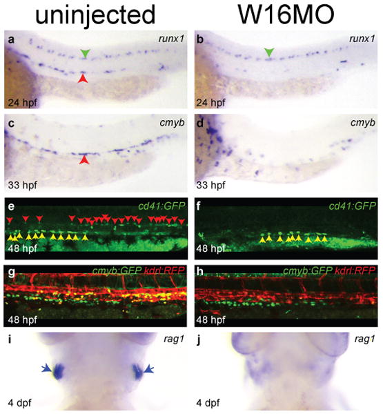
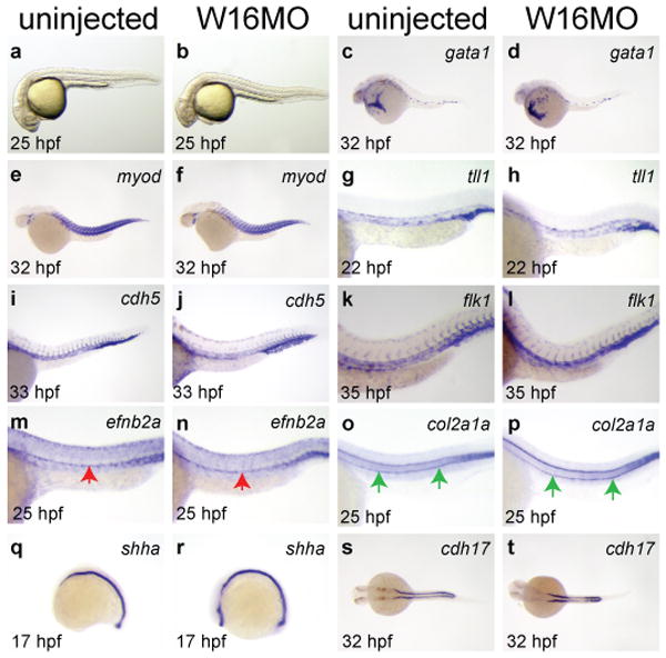
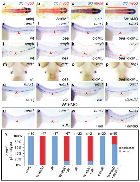
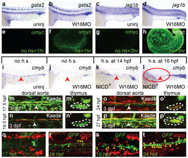
Haematopoietic stem cells (HSCs) are a self-renewing population of cells that continuously replenish all blood and immune cells during the lifetime of an individual. HSCs are used clinically to treat a wide array of diseases, including acute leukaemias and congenital blood disorders, but obtaining suitable numbers of cells and finding immune-compatible donors remain serious problems. These difficulties have led to an interest in the conversion of embryonic stem cells or induced pluripotent stem cells into HSCs, which is not possible using current methodologies. To accomplish this goal, it is critical to understand the native mechanisms involved in the specification of HSCs during embryonic development. Here we demonstrate in zebrafish that Wnt16 controls a novel genetic regulatory network required for HSC specification. Non-canonical signalling by Wnt16 is required for somitic expression of the Notch ligands deltaC (dlc) and deltaD (dld), and these ligands are, in turn, required for the establishment of definitive haematopoiesis. Notch signalling downstream of Dlc and Dld is earlier than, and distinct from, known cell-autonomous requirements for Notch, strongly suggesting that novel Notch-dependent relay signal(s) induce the first HSCs in parallel to other established pathways. Our results demonstrate that somite-specific gene expression is required for the production of haemogenic endothelium.


Conflict of interest statement

The authors declare no competing interests.

Figures

Figure 1
Figure 1. Wnt16 is required for specification of HSCs
Expression of the HSC markers runx1 (a-b) and cmyb (c-d). Fluorescently labelled HSCs in cd41:gfp (e-f) and cmyb:GFP;kdrl:RFP transgenics (g-h). Expression of the lymphocyte marker rag1 (i-j). Embryos are either uninjected (left column) or injected with 5 ng W16MO (right column). Red arrowheads identify the aorta region (a, c) or individual HSCs (e). Green arrowheads identify unaffected runx1+ neurons (a-b). Yellow arrowheads identify unaffected GFP+ multiciliate cells of the pronephros (e-f). Yellow cells are HSCs (g). Blue arrows identify thymic T cells (i). a-h dorsal up, anterior left. i-j ventral views, anterior up.
Figure 2
Figure 2. Wnt16 loss of function phenotype is specific
Uninjected or W16MO-injected embryos in brightfield (a-b) or processed by WISH for tissue specific genes: primitive blood (gata1, c-d), somites (myod, e-f), vasculature (tll1, g-h; cdh5, i-j; flk1, k-l), dorsal aorta (efnb2a, m-n), hypochord (col2a1a, o-p), floorplate and notochord (shha, q-r), or pronephros (cdh17, s-t) at the developmental times indicated. Red arrows, dorsal aorta (m-n); green arrows, hypochord (o-p). Anterior left. a-r, dorsal up. s-t, dorsal views.
Figure 3
Figure 3. Wnt16 acts upstream of Notch ligands Dlc and Dld
Expression of somitic dlc (a-b) and dld (c-d) but not myod (red) is decreased at 17 hpf in W16MO-injected embryos. Red bars indicate somites. Expression of the HSC markers runx1 at 24 hpf (e-h) and cmyb at 36 hpf (i-l) is reduced in dlc mutant (bea) embryos (f, j) or dldMO-injected embryos (g, k), and eliminated in the combined animals (h, l). The lymphocyte marker rag1 at 4.5 dpf is present in wild-type (m), bea (n), and dldMO-injected animals (o), but eliminated in bea embryos injected with dldMO (p). Combined injection of dlc/dld rescues HSCs in wnt16 morphants (q-x). Runx1 at 24 hpf. One group of embryos was uninjected, or injected with the indicated Notch ligand mRNAs alone (q-t). A second group of W16MO-injected embryos was co-injected with Notch ligand mRNAs (u-x). Percentages of embryos displaying the depicted phenotypes (y). a-d flat mounts, anterior left. e-l, q-x close up lateral views, anterior left, dorsal up. m-p ventral head views, anterior up. Red arrowheads indicate the aorta region.
Figure 4
Figure 4. Non-cell-autonomous requirement for Notch in HSC specification
Expression of the Notch target gata2 (a-b) and the Notch ligand jag1b (c-d) in uninjected (a, c) or W16MO-injected (b, d) embryos at 22 hpf. Whole mount immunofluorescence visualization of the myc-tagged NICD at 1 (e-f) and 3 (g-h) hours following either no induction (e, g) or heat-shock induction (f, h). Cmyb expression at 36 hpf in transgenic animals carrying a heat-shock inducible dominant activator of Notch signalling (NICD) in uninjected (i), W16MO-injected (j), W16MO-injected and heat-shock induced at 14 hpf (10-ss; k) or 16 hpf (14-ss; l). Red arrowheads indicate the dorsal aorta region. Red circle where HSCs should normally be expressed (h). Green-to-red photoconvertible Kaede Notch reporter animals were entirely photoconverted at the times indicated at left of each panel pair and imaged at 3 dpf (m-p′). Confocal images of the dorsal aorta (white arrowheads; m-p) and thymus (dashed white outline; m′-p′) reveal photoconverted cells only in the thymi of fish converted at 24 and 27 hpf (o′, p′). Max-projection confocal images of the trunk region of embryos processed by double fluorescence in situ for a Notch reporter GFP transgene (green) and the haematopoietic mesoderm marker etsrp (red) at the times indicated (q-t). Yellow arrowheads indicate double-positive cells (r-t). a-d, i-l close up lateral views of the trunk region, anterior left, dorsal up. e-h, whole embryo views. m-p close up lateral views of the dorsal aorta. m′-p′ single thymic lobes. q-s, dorsal views, anterior left. t, close up lateral aorta view, anterior left, dorsal up.

References

    1. Gering M, Patient R. Notch signalling and haematopoietic stem cell formation during embryogenesis. J Cell Physiol. 2010;222:11–6. - PubMed
    1. Staal FJ, Luis TC. Wnt signaling in hematopoiesis: crucial factors for self-renewal, proliferation, and cell fate decisions. J Cell Biochem. 2010;109:844–9. - PubMed
    1. Angers S, Moon RT. Proximal events in Wnt signal transduction. Nat Rev Mol Cell Biol. 2009;10:468–77. - PubMed
    1. Takada S, et al. Wnt-3a regulates somite and tailbud formation in the mouse embryo. Genes Dev. 1994;8:174–89. - PubMed
    1. Goessling W, et al. Genetic interaction of PGE2 and Wnt signaling regulates developmental specification of stem cells and regeneration. Cell. 2009;136:1136–47. - PMC - PubMed

Publication types

MeSH terms