. 2011 Jun 8;474(7350):230-4.
doi: 10.1038/nature10155.

A function for cyclin D1 in DNA repair uncovered by protein interactome analyses in human cancers

Affiliations

A function for cyclin D1 in DNA repair uncovered by protein interactome analyses in human cancers

Siwanon Jirawatnotai et al. Nature. .

Abstract

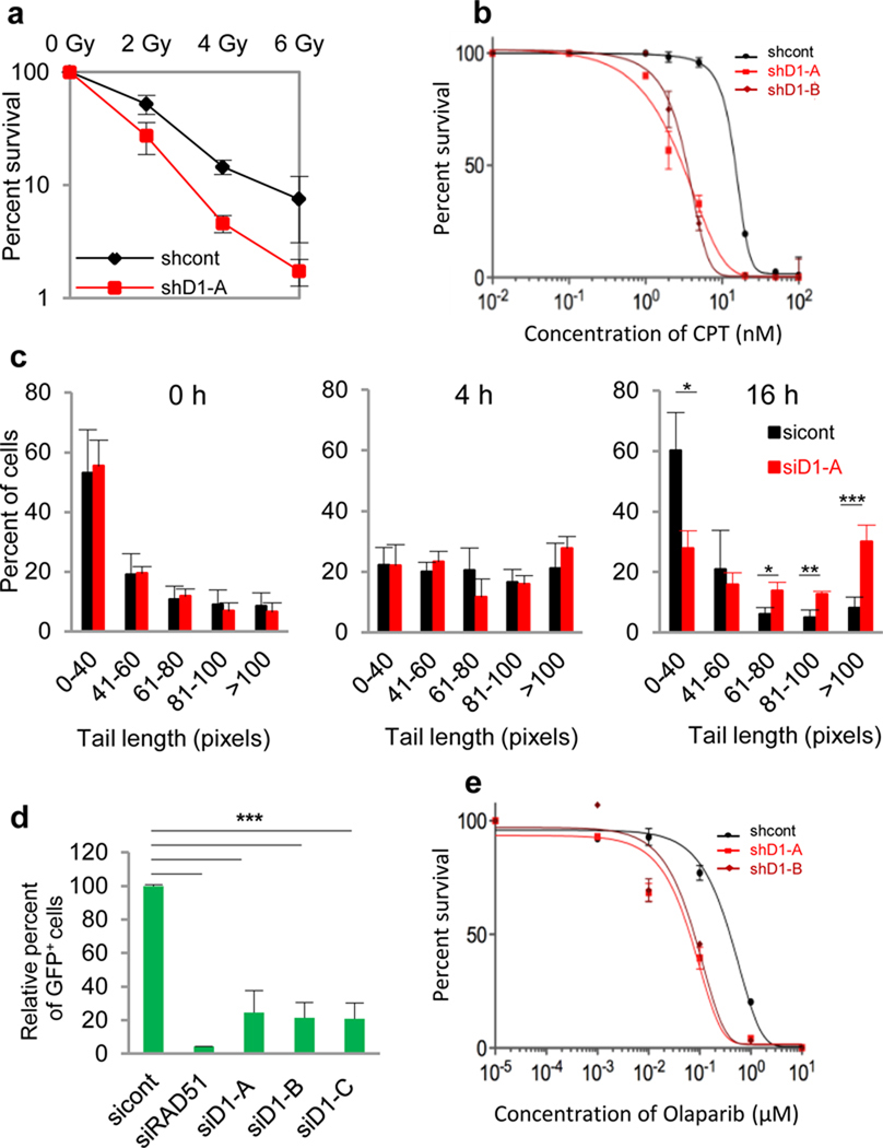
Cyclin D1 is a component of the core cell cycle machinery. Abnormally high levels of cyclin D1 are detected in many human cancer types. To elucidate the molecular functions of cyclin D1 in human cancers, we performed a proteomic screen for cyclin D1 protein partners in several types of human tumours. Analyses of cyclin D1 interactors revealed a network of DNA repair proteins, including RAD51, a recombinase that drives the homologous recombination process. We found that cyclin D1 directly binds RAD51, and that cyclin D1-RAD51 interaction is induced by radiation. Like RAD51, cyclin D1 is recruited to DNA damage sites in a BRCA2-dependent fashion. Reduction of cyclin D1 levels in human cancer cells impaired recruitment of RAD51 to damaged DNA, impeded the homologous recombination-mediated DNA repair, and increased sensitivity of cells to radiation in vitro and in vivo. This effect was seen in cancer cells lacking the retinoblastoma protein, which do not require D-cyclins for proliferation. These findings reveal an unexpected function of a core cell cycle protein in DNA repair and suggest that targeting cyclin D1 may be beneficial also in retinoblastoma-negative cancers which are currently thought to be unaffected by cyclin D1 inhibition.


Figures

Figure 1
Figure 1. Analyses of cyclin D1-interactors identified in human cancers
a, A diagram depicting cyclin D1-interactors, grouped by cell line in which they were detected. Line thickness corresponds to the abundance of peptides for each protein detected in MS. b, Biological process/molecular function enrichment heatmap of cyclin D1-interactors. Each column corresponds to the indicated cell line, rows denote distinct biological process/molecular function. Red color depicts enriched functions, green color – no enrichment. Left panel shows a complete map, right panel an enlarged map of molecular functions/biological processes enriched across all 5 cancer cell lines analyzed.
Figure 2
Figure 2. Impaired DNA repair upon reduction of cyclin D1 levels
a, Colony survival assay of HeLa cells expressing anti-cyclin D1 shRNA (shD1-A) or control shRNA (shcont), after the indicated doses of IR. SF50= 2.15 Gy (shcont), 1.07 Gy (shD1). b, e, Colony survival assays of H2009 cells expressing anti-cyclin D1 shRNAs (shD1-A or shD1-B), or control shRNA (shcont), after exposure to indicated doses of camptothecin (CPT) or AZD2281 (Olaparib). SF50 for camptothecin: 15.1 nM (shcont), 2.8 nM (shD1-A), 3.4 nM (shD1-B); Olaparib: 0.39 µM (shcont), 0.083 µM (shD1-A), 0.065 µM (shD1-B). c, Results of comet assays to measure DNA repair in cyclin D1-depleted HeLa cells (siD1-A) and control cells (sicont). Shown are percentages of cells containing various comet tail lengths at the indicated time-points after IR. d, HR assay in HeLa cells using DR-eGFP-reporter system. sicont, control siRNA; siRAD51, anti-RAD51 siRNA; siD1-A, -B,-C, anti-cyclin D1 siRNAs. *, p ≤ 0.05; **, p ≤ 0.01; ***, p ≤ 0.005; Error bars, standard deviation; n = 3–5.
Figure 3
Figure 3. Functional interaction between cyclin D1 and RAD51
a, Direct binding between GST-cyclin D1 and recombinant RAD51 protein. GST-Synapsin1, GST-Wave1 and GST served as negative controls for RAD51 binding. Upper panel: RAD51 was detected by immunoblotting. Lower panel: input GST-proteins visualized by Ponceau staining. b, Interaction between endogenous cyclin D1 and RAD51, detected by cyclin D1 immunoprecipitation and anti-RAD51 immunoblotting, in indicated cell lines. c, Cyclin D1-RAD51 interaction at different time-points after irradiating HeLa cells. Top panel: cyclin D1 immunoprecipitation - RAD51 immunoblotting. Lower panel: levels of cyclin D1 and RAD51 after IR, determined by western blotting. d, Left panel: Recruitment of RAD51 to I-SceI-induced double-stranded DNA break, gauged by anti-RAD51 ChIP followed by PCR with primers adjacent to DNA damage site. Bars show enrichment around DNA damage site before and after induction of a DNA break. For control, we used anti-E2F4 and non-immune IgG (IgG) ChIP. Right panel: same assay in cells expressing anti-cyclin D1 siRNA. e, Recruitment of cyclin D1 to I-SceI-induced double-stranded DNA break, tested as in d using anti-cyclin D1 ChIP. Bars show enrichment around DNA damage site. f, Co-localization of cyclin D1 and RAD51 at the DNA damage site. Anti-cyclin D1 ChIP was followed by anti-RAD51 re-ChIP and PCR with primers adjacent to double-stranded DNA break. g, Reduction of RAD51 recruitment to DNA damage foci in HeLa cells depleted of cyclin D1 (siD1-A, -C). Percentage of cells displaying a given number of RAD51 foci per nucleus was determined before (0 h) and 8 hours after irradiation. h, In vitro binding assays using the indicated GST-BRCA2 fragments and recombinant cyclin D1. Cyclin D1 was detected by anti-cyclin D1 immunoblotting. Amounts of GST-BRCA2 fragments were verified by Ponceau staining (lower panel). i, Interaction between endogenous cyclin D1 and BRCA2 (B2) in H2009 cells, detected by immunoprecipitation–immunoblotting. j, Recruitment of cyclin D1 to an I-SceI-induced double-stranded DNA break, tested as in e, using cells transfected with independent anti-BRCA2 siRNAs (siBRCA2-A, and -B), or with control siRNA (sicont), Bars show enrichment around DNA damage site. IP, immunoprecipitation; WCL, whole cell lysate; *, p ≤ 0.05; **, p ≤ 0.01; ***, p ≤ 0.005; Error bars, standard deviation; n=3–5.
Figure 4
Figure 4. Increased radiation-sensitivity of tumors with reduced cyclin D1 levels
a, H2009 cells expressing control or anti-cyclin D1 shRNA were injected into nude mice and tumor growth was monitored on the indicated days. Left panel, mice received no radiation. Middle and right panels: at days 5 and 10, tumors were exposed to 2 Gy or 4 Gy of IR. Error bars, standard deviation; **, p ≤ 0.01; ***, p ≤ 0.005. b, Weight of tumors at the end of observation period. Each circle represents an individual tumor, horizontal bars - mean values. N, number of mice analyzed

Comment in

  • DNA repair: Cyclin D1 multitasks.
    Bartek J, Lukas J. Bartek J, et al. Nature. 2011 Jun 8;474(7350):171-2. doi: 10.1038/474171a. Nature. 2011. PMID: 21654798 No abstract available.

References

    1. Sherr CJ, Roberts JM. Living with or without cyclins and cyclin-dependent kinases. Genes Dev. 2004;18:2699–2711. - PubMed
    1. Deshpande A, Sicinski P, Hinds PW. Cyclins and cdks in development and cancer: a perspective. Oncogene. 2005;24:2909–2915. - PubMed
    1. Baumann P, West SC. Role of the human RAD51 protein in homologous recombination and double-stranded-break repair. Trends Biochem Sci. 1998;23:247–251. - PubMed
    1. Bates S, et al. Absence of cyclin D/cdk complexes in cells lacking functional retinoblastoma protein. Oncogene. 1994;9:1633–1640. - PubMed
    1. Lukas J, et al. Retinoblastoma-protein-dependent cell-cycle inhibition by the tumour suppressor p16. Nature. 1995;375:503–506. - PubMed

Publication types

LinkOut - more resources