

Short-term clinical observation of acute retinal pigment epitheliitis using spectral-domain optical coherence tomography
- PMID: 21655052
- PMCID: PMC3102830
- DOI: 10.3341/kjo.2011.25.3.222
Short-term clinical observation of acute retinal pigment epitheliitis using spectral-domain optical coherence tomography
Abstract
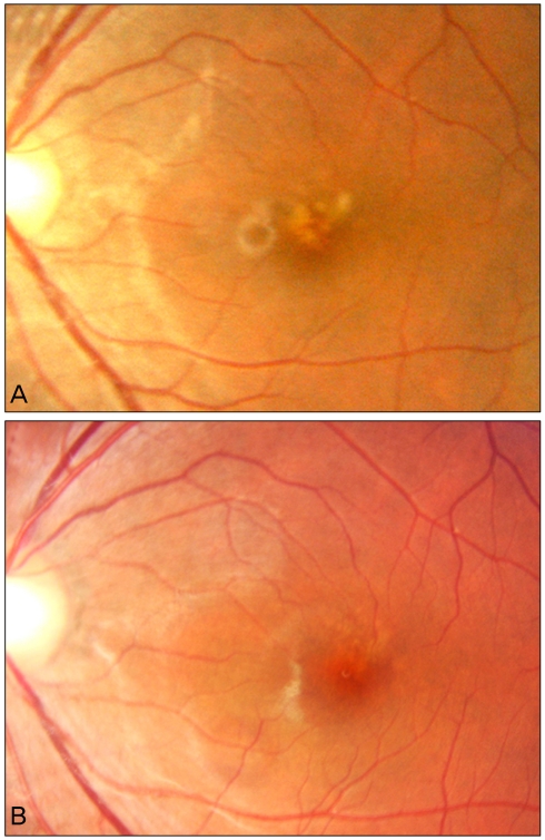
We investigated the case of a young man with blurred vision in his left eye. His visual acuity was slightly decreased, and ophthalmoscopy disclosed a gray-white lesion in the macula. He had no systemic or ocular history. On the visual field test, the threshold sensitivity was decreased in the corresponding region. Spectral domain optical coherence tomography (OCT) demonstrated a disruption in the photoreceptor inner and outer segment (IS/OS) junction and undulation of the retinal pigment epithelium (RPE) with backscattering. We re-examined the patient after two weeks and after three months without any treatment. Visual acuity and visual field results were gradually normalized, and OCT demonstrated the recovery of continuity in the photoreceptor IS/OS junction, as well as decreased RPE irregularity with minimal backscattering. We used spectral domain OCT instead of time domain OCT (OCT3) so that we could provide better image resolution of the acute retinal pigment epitheliitis (ARPE). Finally, we observed recovery of the functional and anatomical changes in the ARPE patient with a resolution of the condition within three months following the initial examination, using OCT and visual field tests.
Keywords: Acute retinal pigment epitheliitis; Spectral-domain optical coherence tomography.
Conflict of interest statement
No potential conflict of interest relevant to this article was reported.
Figures

References
-
- Krill AE, Deutman AF. Acute retinal pigment epitheliitus. Am J Ophthalmol. 1972;74:193–205. - PubMed
-
- Chittum ME, Kalina RE. Acute retinal pigment epitheliitis. Ophthalmology. 1987;94:1114–1119. - PubMed
-
- Hsu J, Fineman MS, Kaiser RS. Optical coherence tomography findings in acute retinal pigment epitheliitis. Am J Ophthalmol. 2007;143:163–165. - PubMed
-
- Quillen DA, Davis JB, Gottlieb JL, et al. The white dot syndromes. Am J Ophthalmol. 2004;137:538–550. - PubMed
-
- Li D, Kishi S. Restored photoreceptor outer segment damage in multiple evanescent white dot syndrome. Ophthalmology. 2009;116:762–770. - PubMed
Publication types
MeSH terms
LinkOut - more resources
Full Text Sources
