. 2011;6(5):e20513.
doi: 10.1371/journal.pone.0020513. Epub 2011 May 31.

Ecdysteroid-dependent expression of the tweedle and peroxidase genes during adult cuticle formation in the honey bee, Apis mellifera

Affiliations

Ecdysteroid-dependent expression of the tweedle and peroxidase genes during adult cuticle formation in the honey bee, Apis mellifera

Michelle P M Soares et al. PLoS One. 2011.

Abstract

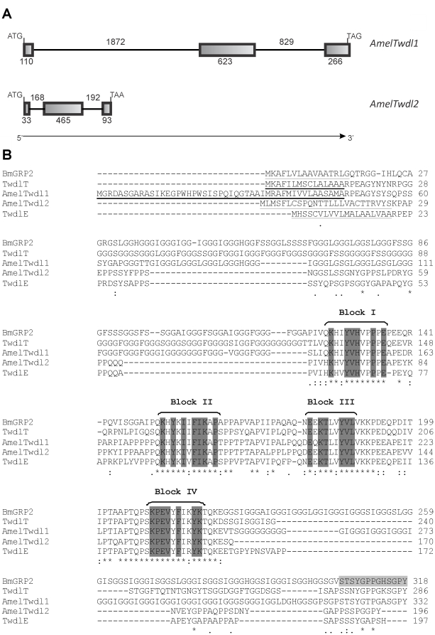
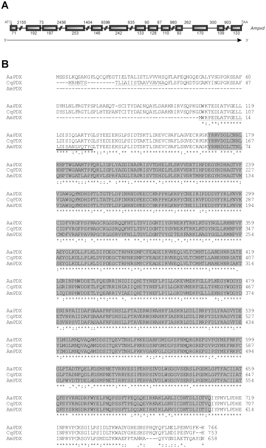
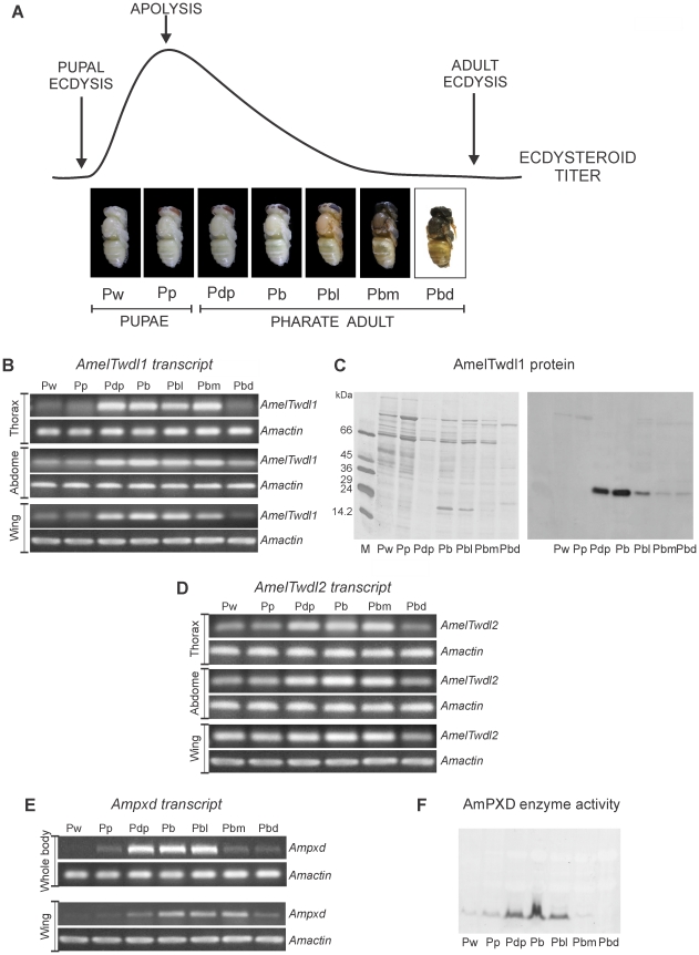
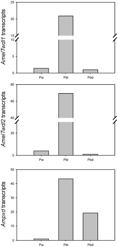
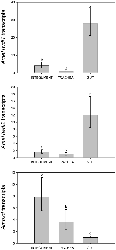
Cuticle renewal is a complex biological process that depends on the cross talk between hormone levels and gene expression. This study characterized the expression of two genes encoding cuticle proteins sharing the four conserved amino acid blocks of the Tweedle family, AmelTwdl1 and AmelTwdl2, and a gene encoding a cuticle peroxidase containing the Animal haem peroxidase domain, Ampxd, in the honey bee. Gene sequencing and annotation validated the formerly predicted tweedle genes, and revealed a novel gene, Ampxd, in the honey bee genome. Expression of these genes was studied in the context of the ecdysteroid-coordinated pupal-to-adult molt, and in different tissues. Higher transcript levels were detected in the integument after the ecdysteroid peak that induces apolysis, coinciding with the synthesis and deposition of the adult exoskeleton and its early differentiation. The effect of this hormone was confirmed in vivo by tying a ligature between the thorax and abdomen of early pupae to prevent the abdominal integument from coming in contact with ecdysteroids released from the prothoracic gland. This procedure impaired the natural increase in transcript levels in the abdominal integument. Both tweedle genes were expressed at higher levels in the empty gut than in the thoracic integument and trachea of pharate adults. In contrast, Ampxd transcripts were found in higher levels in the thoracic integument and trachea than in the gut. Together, the data strongly suggest that these three genes play roles in ecdysteroid-dependent exoskeleton construction and differentiation and also point to a possible role for the two tweedle genes in the formation of the cuticle (peritrophic membrane) that internally lines the gut.


Conflict of interest statement

Competing Interests: The authors have declared that no competing interests exist.

Figures

Figure 1
Figure 1. Tweedle: gene structures and alignment of insect tweedle proteins.
(A) Schematic representation of the AmelTwdl1 and AmelTwdl2 genes. Initiation and termination codons are indicated at the left and right of the figure, respectively. Exons and introns are indicated by boxes and lines, respectively, and the number of nucleotides is shown. The direction of transcription is indicated by an arrow. (B) Alignment (ClustalW 2) of AmelTwdl1 (ACJ38118.1) and AmelTwdl2 (ADK73965.2) amino acid sequences with other Tweedle protein sequences from B. mori, BmGRP2 (BAE06189.1), and D. melanogaster, TwdlT (AAF56656.1) and TwdlE (AAF52571.2). The four conserved blocks of amino acids are marked in dark gray. The signal peptide region is underlined in all sequences. In light gray is a region of BmGRP2 sequence that was used by Zhong et al. (see ref. [40]) to synthesize a peptide for antibody production. This antibody recognized the AmelTwdl1 protein (see Fig. 3 and corresponding text in Results section). Asterisks, colons and dots represent identical amino acid residues, strong- and weak-conservative substitutions, respectively.
Figure 2
Figure 2. Peroxidase: gene structure and alignment of insect peroxidase proteins.
(A) Schematic representation of the peroxidase gene from A. mellifera. Initiation and termination codons are indicated, as well as exons (boxes) and introns (lines). The number of nucleotides is shown, and the direction of transcription is indicated by an arrow. (B) Alignment (ClustalW 2) of peroxidase sequences from A. mellifera (AmPXD, ADE45321.2), Culex quinquefasciatus (CqPXD, EDS26535.1) and Aedes aegypti (AaPXD, EAT46477.1). The signal peptide region was underlined in the A. mellifera and C. quinquefasciatus sequences. The region containing the Animal haem peroxidase domain (pfam03098) is marked in grey. Asterisks, colons and dots represent identical amino acid residues, strong- and weak-conservative substitutions, respectively.
Figure 3
Figure 3. Expression of AmelTwdl1, AmelTwdl2 and Ampxd genes during the pupal-to-adult development coordinated by ecdysteroid titer.
(A) Hemolymph ecdysteroid titer redrawn from Pinto et al. (see ref. [59]), and the successive developmental phases in the interval between the pupal and adult ecdyses: Pw and Pp are early and late pupae; Pdp, Pb, Pbl, Pbm and Pbd are the successive pharate adult phases. (B) Transcriptional profile of AmelTwdl1 in the thoracic, abdominal and wing integument. (C) Developmental profile of thoracic integument proteins stained with Coomassie Brillant Blue (at the left) and the AmeTwdl1 protein (at the right) as detected using Western blot and an antibody against a cuticle protein from B. mori (BmGRP2). (D) Transcriptional profile of AmelTwdl2 in the thoracic, abdominal and wing integument. (E) Transcriptional profile of Ampxd in whole body extracts and wings. (F) AmPXD enzyme activity detected in thoracic integument samples using electrophoresis in polyacrylamide gels stained with a specific substrate. Transcript abundance was investigated by semiquantitative RT-PCR followed by electrophoresis of the amplified cDNAs in ethidium bromide-stained agarose gels. An A. mellifera actin gene, Amactin, was used as endogenous control.
Figure 4
Figure 4. Relative quantification of AmelTwdl1, AmelTwdl2 and Ampxd transcripts in the thoracic integument of pupae (Pw phase) and pharate adults (Pbl and Pbd phases).
The Amrp49 gene was used as endogenous control in real-time RT-PCR assays, and the relative amount of transcripts is given by 2-ΔΔC T. Each column represents the mean of two independent integument samples.
Figure 5
Figure 5. Relative quantification of AmelTwdl1, AmelTwd2 and Ampxd transcripts in different tissues of the Pbl pharate adult.
The Amrp49 gene was used as endogenous control in real-time RT-PCR assays. The relative amount of transcripts is given by 2-ΔΔC T. Columns and bars represent means±SE of three independent samples prepared with each tissue. AmelTwdl1 transcript levels were significantly different among the tested tissues, with a higher expression in the gut (p = 0.002). Similarly, AmelTwdl2 showed a significantly higher expression in the gut than in integument and trachea (p = 0.010). In contrast, Ampxd showed a higher expression in integument than in the other tissues (p = 0.001). Statistical analysis was carried out with Jandel SigmaStat 3.1 software (Jandel Corporation, San Rafael, CA, USA). One Way Anova; post-hoc comparisons using Holm Sidak test p<0.05. Different letters above columns indicate statistical difference.
Figure 6
Figure 6. Abundance of AmelTwdl1, AmelTwdl2 and Ampxd transcripts in the integument of ligated (L) abdomens compared to non-ligated controls (NL).
Transcript levels were investigated at days (d) 1, 2, 3, 4, 5 and 6 after the abdominal ligature of newly ecdysed pupae. Transcript levels were assessed by semiquantitative RT-PCR assays followed by electrophoresis of the amplified cDNAs in ethidium bromide stained agarose gels. The Amactin gene was used as endogenous control.

References

    1. Hepburn HR. Structure of the integument. In: Kerkut GA, Gilbert LI, editors. Comprehensive Insect Physiology, Biochemistry and Pharmacology. Oxford: Pergamon Press; 1985. pp. 1–58.
    1. Gorman MJ, Arakane Y. Tyrosine hydroxylase is required for cuticle sclerotization and pigmentation in Tribolium castaneum. Insect Biochemistry and Molecular Biology. 2010;40:267–273. - PMC - PubMed
    1. Hiruma K, Carter MS, Riddiford LM. Characterization of the dopa decarboxylase gene of Manduca sexta and its suppression by 20-Hydroxyecdysone. Developmental Biology. 1995;169:195–209. - PubMed
    1. Hiruma K, Riddiford LM. The molecular mechanisms of cuticular melanization: The ecdysone cascade leading to dopa decarboxylase expression in Manduca sexta. Insect Biochemistry and Molecular Biology. 2009;39:245–253. - PubMed
    1. Arakane Y, Dixit R, Begum K, Park Y, Specht CA, et al. Analysis of functions of the chitin deacetylase gene family in Tribolium castaneum. Insect Biochem Mol Biol. 2009;39:355–365. - PubMed

Publication types