. 2011;6(5):e20345.
doi: 10.1371/journal.pone.0020345. Epub 2011 May 31.

The membrane bound LRR lipoprotein Slr, and the cell wall-anchored M1 protein from Streptococcus pyogenes both interact with type I collagen

Affiliations

The membrane bound LRR lipoprotein Slr, and the cell wall-anchored M1 protein from Streptococcus pyogenes both interact with type I collagen

Marta Bober et al. PLoS One. 2011.

Abstract

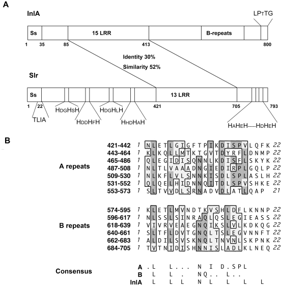
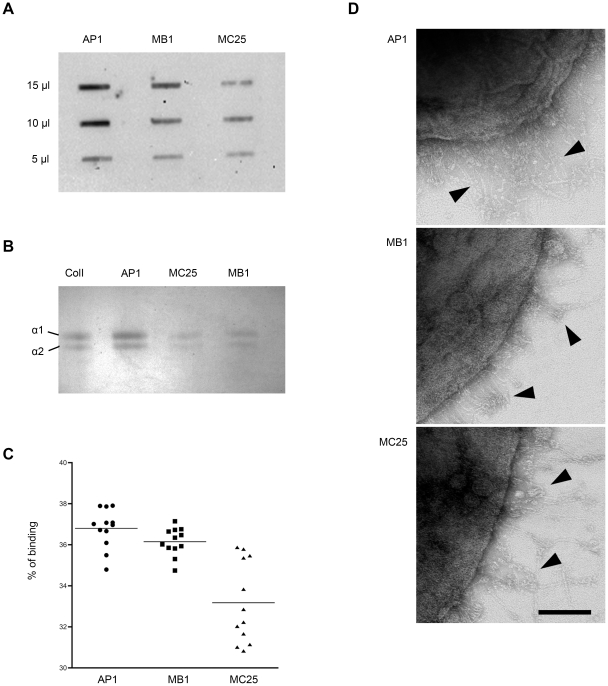
Streptococcus pyogenes is an important human pathogen and surface structures allow it to adhere to, colonize and invade the human host. Proteins containing leucine rich repeats (LRR) have been identified in mammals, viruses, archaea and several bacterial species. The LRRs are often involved in protein-protein interaction, are typically 20-30 amino acids long and the defining feature of the LRR motif is an 11-residue sequence LxxLxLxxNxL (x being any amino acid). The streptococcal leucine rich (Slr) protein is a hypothetical lipoprotein that has been shown to be involved in virulence, but at present no ligands for Slr have been identified. We could establish that Slr is a membrane attached horseshoe shaped lipoprotein by homology modeling, signal peptidase II inhibition, electron microscopy (of bacteria and purified protein) and immunoblotting. Based on our previous knowledge of LRR proteins we hypothesized that Slr could mediate binding to collagen. We could show by surface plasmon resonance that recombinant Slr and purified M1 protein bind with high affinity to collagen I. Isogenic slr mutant strain (MB1) and emm1 mutant strain (MC25) had reduced binding to collagen type I as shown by slot blot and surface plasmon resonance. Electron microscopy using gold labeled Slr showed multiple binding sites to collagen I, both to the monomeric and the fibrillar structure, and most binding occurred in the overlap region of the collagen I fibril. In conclusion, we show that Slr is an abundant membrane bound lipoprotein that is co-expressed on the surface with M1, and that both these proteins are involved in recruiting collagen type I to the bacterial surface. This underlines the importance of S. pyogenes interaction with extracellular matrix molecules, especially since both Slr and M1 have been shown to be virulence factors.


Conflict of interest statement

Competing Interests: Hansa Medical AB is one of the funders of this study. This does not alter the authors‚ adherence to all the PLoS ONE policies on sharing data and materials.

Figures

Figure 1
Figure 1. Characteristics of the Slr protein.
A: Schematic comparison of the InlA from L. monocytogenes and Slr. The identity and similarity between the two LRR regions are stated. The cell wall anchoring motif for InlA and the signal peptide II recognition motif for Slr are indicated as well as the histidine triad motifs. B: Sequence alignments of the two groups of LRRs (A and B repeats) in Slr and comparison of the LRR consensus sequences of Slr and InlA.
Figure 2
Figure 2. Visualization and confirmation of the LRR lipoprotein Slr.
A: Slr protein visualized by electron microscopy showing the typical horseshoe shape of a LRR protein. Scale bar  = 25 nm. B: a homology model of Slr using InlA as the template. The 11 LRRs forming β-sheets in yellow, α−helices in blue, and loops in turquoise. C: SDS-PAGE analysis of recombinantly expressed GST-Slr and Slr with the GST tag cleaved off. D: Western blot analysis, using anti-Slr antibodies, of bacterial cell extracts from AP1 bacteria grown in the presence of increasing amounts of the signal peptidase II inhibitor globomycin as indicated.
Figure 3
Figure 3. Slr gene disruption, mutant strain analysis and protein localization by electron microscopy.
A: Schematic representation of the mutagenesis strategy used to disrupt slr in S. pyogenes strain AP1. B: SDS-PAGE and Western blot analysis, using antibodies against Slr and M1, of bacterial cell extracts of the mutant strains MB1 and MC25, and wild type bacteria AP1. C: Wild type strain AP1, mutant strain MB1 and mutant strain MC25 incubated with gold labeled F(ab')2 fragments of anti-Slr (small gold particles) and anti-M1 (big gold particles) IgG. The dots represent binding of the F(ab')2 fragments to the Slr and M1 protein on the bacterial surface. Arrowheads without arrows indicate binding of anti-Slr antibodies, and complete arrows indicate binding of anti-M1 antibodies in all three panels. Scale bar  = 100 nm.
Figure 4
Figure 4. Growth curve and expression pattern of Slr and M1 protein in S. pyogenes strain AP1.
A: Growth curve of S. pyogenes wild type strain AP1 growth in THY medium over time. B: Western blot analysis of Slr and M1 protein expression in strain AP1 over 9 hours of growth.
Figure 5
Figure 5. Absorption of collagen type I to S. pyogenes strains.
A: After incubation of mutant strains MB1, MC25 and wild type AP1 strain with collagen I, low pH eluates from bacteria were immobilized on a PVDF membrane and detected using anti-collagen antibodies. The amount of collagen I eluate applied on the membrane is indicated to the left. B: The bacterial eluates described in 4A were separated on SDS-PAGE and stained with Coomassie Brilliant Blue. Visible are the α1 (134 kDa) and α2 (130 kDa) chains indicated with arrows. C: Binding in percentage of 125I labeled collagen I to the bacterial strains AP1, MB1 and MC25. Experiments were performed in quadruples at three independent time points. D: Interaction between S. pyogenes strains and monomeric collagen. Collagen I was incubated with AP1, MB1 and MC25 and visualized by electron microscopy. Arrows point to collagen I monomers. Scale bar  = 100 nm.
Figure 6
Figure 6. Binding of Slr to immobilized collagen I.
A: Collagen I denatured with 4 M guanidine hydrochloride (+) and non-denatured collagen I (−) were applied to a PVDF membrane and incubated with radiolabeled Slr. The amount of collagen is indicated to the right. B: Binding of gold labeled Slr (the dots) and collagen I using electron microscopy. The dots are bound to C- and N terminal as well as 70 respectively 100 nm further in on the monomeric collagen I (panel I). The short span between light strands represents the overlap region, whiles the dark span represents the gap region on a collagen I fibril. Binding can be observed in both regions, but is mostly concentrated to the overlap region (panel II). Scale bar  = 100 nm.
Figure 7
Figure 7. Surface plasmon resonance interaction analysis.
The binding of Slr and M1 protein, the wild type strain AP1, and mutant strains MB1 and MC25 strains to immobilized collagen I was investigated using surface plasmon resonance. Binding curves are displayed and, the association (Ka) and dissociation (Kd) rate constants were determined. A–B: Slr and M1 proteins were diluted 2-fold in running buffer and injected over the collagen surface starting at 500 nM and 35 µl/min. The proteins displayed a dose-dependent binding to collagen I with a KD of 12 nM for Slr and 54 nM for M1 protein. C–E: Bacterial cell suspensions were diluted 2-fold into PBS for injections sequences starting from 2×10⁹ cfu/ml solution.

References

    1. Cunningham MW. Pathogenesis of group A streptococcal infections. Clin Microbiol Rev. 2000;13:470–511. - PMC - PubMed
    1. Dinkla K, Rohde M, Jansen WT, Kaplan EL, Chhatwal GS, et al. Rheumatic fever-associated Streptococcus pyogenes isolates aggregate collagen. J Clin Invest. 2003;111:1905–1912. - PMC - PubMed
    1. Åkesson P, Schmidt KH, Cooney J, Björck L. M1 protein and protein H: IgGFc- and albumin-binding streptococcal surface proteins encoded by adjacent genes. Biochem J. 1994;300(Pt 3):877–886. - PMC - PubMed
    1. Kreikemeyer B, Nakata M, Oehmcke S, Gschwendtner C, Normann J, et al. Streptococcus pyogenes collagen type I-binding Cpa surface protein. Expression profile, binding characteristics, biological functions, and potential clinical impact. J Biol Chem. 2005;280:33228–33239. - PubMed
    1. Jin MS, Lee JO. Structures of TLR-ligand complexes. Curr Opin Immunol. 2008;20:414–419. - PubMed

Publication types

MeSH terms