. 2011;6(5):e20495.
doi: 10.1371/journal.pone.0020495. Epub 2011 May 31.

Immunolocalization of influenza A virus and markers of inflammation in the human Parkinson's disease brain

Affiliations

Immunolocalization of influenza A virus and markers of inflammation in the human Parkinson's disease brain

Troy T Rohn et al. PLoS One. 2011.

Abstract

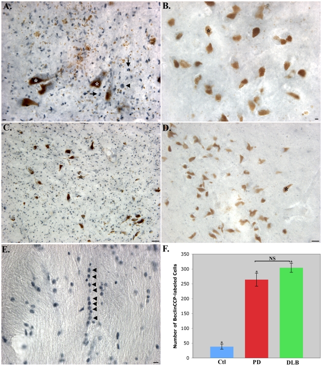
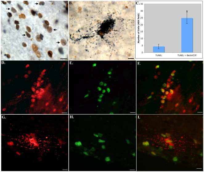
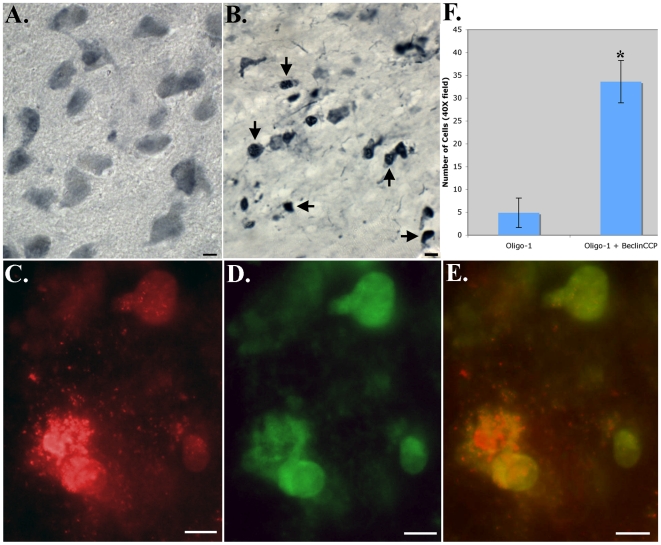
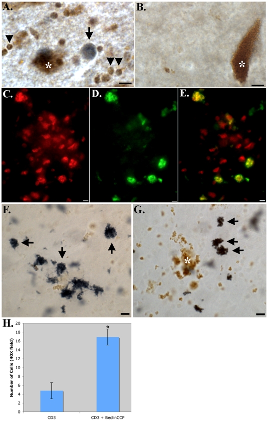
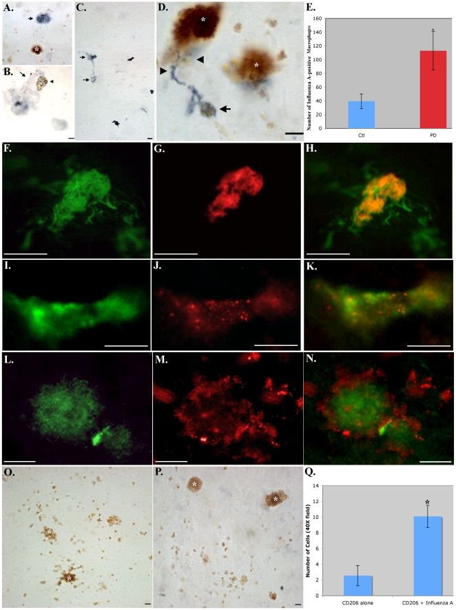
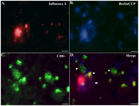
Although much is known regarding the molecular mechanisms leading to neuronal cell loss in Parkinson's disease (PD), the initiating event has not been identified. Prevailing theories including a chemical insult or infectious agent have been postulated as possible triggers, leading to neuroinflammation. We present immunohistochemical data indicating the presence of influenza A virus within the substantia nigra pars compacta (SNpc) from postmortem PD brain sections. Influenza A virus labeling was identified within neuromelanin granules as well as on tissue macrophages in the SNpc. Further supporting a role for neuroinflammation in PD was the identification of T-lymphocytes that colocalized with an antibody to caspase-cleaved Beclin-1 within the SNpc. The presence of influenza A virus together with macrophages and T-lymphocytes may contribute to the neuroinflammation associated with this disease.


Conflict of interest statement

Competing Interests: The authors have declared that no competing interests exist.

Figures

Figure 1
Figure 1. Caspase-cleaved Beclin-1 in Parkinson's and dementia with Lewy body disease.
(A–D): High (A and B) and low magnification (C and D), of representative single-labeling (blue) from a Parkinson's cases utilizing the BeclinCCP antibody illustrating staining of numerous small cells (<10 µm) in the SNpc (arrows, Fig. 1A) along with the staining of a single Lewy body (arrowhead, Fig. 1A). Comparative staining in representative age-matched control cases showing a general lack of staining with the BeclinCCP antibody (B and D). Brown structures (A–D) represent neuromelanin (asterisks), typical of neurons in the SNpc. (E): Representative labeling in a DLB case illustrating the “pearls-on-a-string” labeling with BeclinCCP in white matter within the SN (E, arrowheads). (F): Quantification of the number of BeclinCCP-positive cells within the SNpc for age-matched control cases (blue bar), PD cases, (red bar) and DLB cases (green bar). Results indicated a significant increase in the number of BeclinCCP-positive cells in both PD and DLB over control cases. Data represent the average (±S.E.M.) of three different fields taken with a 40× objective from five different cases. NS = no significant difference between PD and DLB cases (p = 0.331). *PD indicates significant difference between PD and control cases (p = 0.0005), and *DLB indicates significant difference between DLB and control cases (p = 0.0001). Scale bars are 10 µm in A, B, and E and 50 µm in C and D.
Figure 2
Figure 2. Co-localization of the BeclinCCP antibody with TUNEL labeling, a marker for apoptosis.
(A and B): Representative bright-field double-labeling utilizing the BeclinCCP antibody (blue) and TUNEL (DAB, brown) revealed the co-localization within small cells (arrows, A) and within a degenerating astrocyte (B) in SNpc of representative Parkinson's cases. (D–I): Representative immunofluorescence double-labeling within the SN utilizing BeclinCCP (red) and TUNEL (green) in a representative PD case (D–F) or DLB (G–I). Panel G illustrates the staining of a degenerating astrocyte that co-localized with TUNEL (Panel I). (C): Panel C depicts quantitative immunofluorescence analysis indicating that approximately 85% of TUNEL-positive cells co-localized with the BeclinCCP antibody (p = 1.38×10⁻⁵). Scale bars are 10 µm.
Figure 3
Figure 3. Degenerating oligodendrocytes co-localize with caspase-cleaved Beclin-1 in PD.
(A and B): Representative labeling in control or PD cases utilizing mAB anti-Olig1 indicated staining of well-defined, healthy oligodendrocytes in age-matched control cases (A), as compared to PD cases where labeling was identified on oligodendrocytes exhibiting shrunken cell bodies (B). (C–E): Immunofluorescence double labeling in a representative PD case indicated the co-localization of the BeclinCCP antibody (red, C) with anti-Olig1 (green, D). Panel E displays the overlap image for both antibodies indicating the labeling of the BeclinCCP antibody within degenerating oligodendrocytes (yellow, E). F): Panel F depicts quantitative analysis indicating that approximately 87% of anti-Olig1-positive cells co-localized with the BeclinCCP antibody (p = 1.46×10⁻⁶). All scale bars are 10 µm.
Figure 4
Figure 4. Caspase-cleaved Beclin-1 within T-lymphocytes in the PD brain.
(A and B): Bright-field double labeling in the SNpc of a representative PD case (A) or control (B) illustrating co-localization of the BeclinCCP antibody (blue) with mAB CD3, a marker for T-lymphocytes (brown). Note the size and co-localization of both markers in cells (arrowheads, A), and in addition the single labeling of BeclinCCP within an apparent Lewy body (arrow, A). There was a general lack of labeling of both markers in control cases (B). Brown structures (A, B, F, and G) represent neuromelanin (asterisks), typical of neurons in the SNpc. (C–E): Representative immunofluorescence double labeling employing the BeclinCCP antibody (red, C) and mAB CD3 (green, D) in the SNpc of a representative PD case, with the overlapped image for both markers (yellow, E). (F and G): Representative staining in the SNpc of a PD case with anti-CD4 in (F) and anti-CD8 (G), markers for helper T-lymphocytes and cytotoxic T-lymphocytes, respectively (arrows). (H): Panel H depicts quantitative analysis indicating that approximately 78% of CD3-positive cells co-localized with the BeclinCCP antibody (p = 7.08×10⁻⁵). Scale bars represent 10 µm.
Figure 5
Figure 5. Evidence for influenza A virus on macrophages within the PD brain.
(A–C): Representative staining (blue) within the SNpc of the PD brain utilizing mAB anti-influenza A virus antibody (35–481) depicting staining within large globular cells consistent with a morphology of macrophages (arrows, A–C). Panel B depicts staining within a macrophage-like cell extending an apparent pseudopod engulfing a smaller circular structure containing neuromelanin with punctate blue labeling (arrowhead, B). (D): Representative anti-influenza A virus staining (blue) in a control case identifying a single macrophage-like cell (arrowheads) surrounding a particle of neuromelanin material (arrow). (E): Quantitative analysis of the number of influenza A-positive macrophages labeled in Ctl (n = 5) or PD cases (n = 4) indicated a significant increase in PD cases (±S.D., p-value = 0.01). (F–K): Double labeling immunofluorescence in the SNpc of representative PD cases utilizing mAB anti-CD206, a macrophage marker (green, F and I), along with anti-influenza A virus labeling (35–481) (red, G and J) with the overlap image (yellow/orange, H and K). Two types of labeling were observed: In rounder macrophages (F), influenza A staining was more localized and homogenous (G). In perivascular regions, macrophages were more elongated (I) and influenza A labeling was clearly punctated in appearance (J). (L–N): Identical to Panels F–K, except with the use of a different antibody marker to both macrophages (CD14, green, L) and influenza A virus (35–483, red, M), with overlap image shown in Panel N. Punctate labeling of influenza A virus was observed on the surface of macrophages. (O): Control experiment showing lack of influenza A blue labeling in the absence of primary antibody. (P): Representative staining in a PD case utilizing an antibody against influenza B, indicating a general lack of blue labeling with this antibody. (Q): Panel Q depicts quantitative analysis indicating that approximately 80% of anti-CD206-positive cells co-localized with the anti-influenza A virus antibody (p = 4.55×10⁻⁶). Brown structures (A, D, O, and P) represent neuromelanin (asterisks), typical of neurons in the SNpc. All scale bars are 10 µm.
Figure 6
Figure 6. Influenza A-positive macrophages co-localize with caspase-cleaved Beclin-1 and are surrounded by cytotoxic T-lymphocytes.
Representative immunofluorescence triple labeling in a PD case was undertaken using three different antibodies: (A) anti-influenza A (red), (B) BeclinCCP (blue), and (C) anti-CD8 (green). (D): Panel D displays the overlapped image for all three antibodies indicating the presence of influenza A viral proteins on an apparent macrophage displaying diffuse BeclinCCP labeling (arrow, D). In addition, numerous CD8+ cells are in close proximity to the macrophage and some of these cells demonstrated colocalization with the influenza A virus antibody (arrowheads, D). All scale bars are 10 µm.

Similar articles

Cited by

References

    1. Reichmann H. Clinical criteria for the diagnosis of Parkinson's disease. Neurodegener Dis. 2010;7:284–290. - PubMed
    1. Dickson DW. Alpha-synuclein and the Lewy body disorders. Curr Opin Neurol. 2001;14:423–432. - PubMed
    1. Ravenholt RT, Foege WH. 1918 influenza, encephalitis lethargica, parkinsonism. Lancet. 1982;2:860–864. - PubMed
    1. Kumar A, Calne SM, Schulzer M, Mak E, Wszolek Z, et al. Clustering of Parkinson disease: shared cause or coincidence? Arch Neurol. 2004;61:1057–1060. - PubMed
    1. Goldsmith JR, Herishanu YO, Podgaietski M, Kordysh E. Dynamics of parkinsonism-Parkinson's disease in residents of adjacent kibbutzim in Israel's Negev. Environ Res. 1997;73:156–161. - PubMed

Publication types