. 2011;6(5):e20108.
doi: 10.1371/journal.pone.0020108. Epub 2011 May 31.

Remodeling of monoplanar Purkinje cell dendrites during cerebellar circuit formation

Affiliations

Remodeling of monoplanar Purkinje cell dendrites during cerebellar circuit formation

Megumi Kaneko et al. PLoS One. 2011.

Abstract

Dendrite arborization patterns are critical determinants of neuronal connectivity and integration. Planar and highly branched dendrites of the cerebellar Purkinje cell receive specific topographical projections from two major afferent pathways; a single climbing fiber axon from the inferior olive that extend along Purkinje dendrites, and parallel fiber axons of granule cells that contact vertically to the plane of dendrites. It has been believed that murine Purkinje cell dendrites extend in a single parasagittal plane in the molecular layer after the cell polarity is determined during the early postnatal development. By three-dimensional confocal analysis of growing Purkinje cells, we observed that mouse Purkinje cells underwent dynamic dendritic remodeling during circuit maturation in the third postnatal week. After dendrites were polarized and flattened in the early second postnatal week, dendritic arbors gradually expanded in multiple sagittal planes in the molecular layer by intensive growth and branching by the third postnatal week. Dendrites then became confined to a single plane in the fourth postnatal week. Multiplanar Purkinje cells in the third week were often associated by ectopic climbing fibers innervating nearby Purkinje cells in distinct sagittal planes. The mature monoplanar arborization was disrupted in mutant mice with abnormal Purkinje cell connectivity and motor discoordination. The dendrite remodeling was also impaired by pharmacological disruption of normal afferent activity during the second or third postnatal week. Our results suggest that the monoplanar arborization of Purkinje cells is coupled with functional development of the cerebellar circuitry.


Conflict of interest statement

Competing Interests: The authors have declared that no competing interests exist.

Figures

Figure 1
Figure 1. Confocal analysis of dendrite arborization in developing Purkinje cells.
A: Sagittal (left) and coronal (right) views of developing Purkinje cells labeled with adeno-associated virus (AAV)-derived GFP. Three-dimensional images were compiled from 20–50 z-serial sections taken at 1 µm intervals. Dendritic processes at P7 orient randomly in the molecular layer. At P9 and thereafter, Purkinje cells bear a single to a few primary stem dendrites which extend branches along the sagittal axis of the molecular layer. B: Confocal and graphic images of typical Purkinje cells at P18. Sagittal (left panels) and coronal (right panels) views are shown. Some dendrites extrude from the sagittal plane filled by main arbors, and further branch in distinct parallel sagittal planes (pseudocolored in yellow and blue in graphic images; see also Video S1). C: The P22 Purkinje cell arborizes dendrites in a single sagittal plane (see also Video S2). Scale bars: 20 µm.
Figure 2
Figure 2. Remodeling of Purkinje dendrites in the third postnatal week.
A: Confocal and graphic images of dendrites in a P35 Purkinje cell. Some branches extrude from the main sagittal plane and overpass other branches (magenta circles). Scale bar: 10 µm. B–F: Quantitative analyses of dendrite development in Purkinje cells in lobules IX and X. The number (B) and total length (C) of branches per cell rapidly increase over the first 18 postnatal days. Both the number and length significantly decrease between P18 and P22, and further increase into adulthood. D: Overpassing branches per cell peak at P18, sharply decrease by P22 and plateau thereafter. E: The mean length of each dendritic branch decreases between P7 and P9, constantly increases until P35 and then slightly decreased by P50. F: The total sagittal-sectional area of the molecular layer covered by the dendrite increases until P35 with a plateau between P18 and P22. Purkinje cells per data point in CG: P7, P14–P50, n = 14; P9, n = 20. Error bars indicate s.e.m. G: Histogram showing the developmental change in percentages of multiplanar Purkinje cells. The percentage of multiplanar Purkinje cells peaks at P18 in both early and late maturing lobules (lobules IX, X and lobules III–VIII, respectively). The number of cells analyzed is indicated in parentheses.
Figure 3
Figure 3. Multiple CF associations to multiplanar dendrites of Purkinje cells.
A: Triple fluorescence for AAV-GFP-infected Purkinje cells (pseudocolored in blue), BDA-labeled CFs (magenta) and vesicular glutamate transporter VGluT2 (green). A CF targets the soma of a Purkinje cell and ramifies into several tendril fibers that run along thick stem dendrites (low magnification views on left). The varicose swellings along the CF overlap with VGluT2-positive puncta indicating CF terminals (high magnification views on right). B: Confocal (left) and graphic (right) images of CFs and a multiplanar Purkinje cell at P18. Cell-1 and Cell-2 are associated by at least 2 and 3 CFs, respectively. The main ascending fibers are indicated by arrowheads in confocal images and by yellow in graphic images. The minor CFs apposing distal dendrites (asterisks in confocal images; light and dark pink in graphic images) innervate the soma of nearby Purkinje cells (circles in confocal images). Dendrites in minor planes are pseudocolored in blue and green in graphic images. Coronal views of graphic images on right indicate that both dendrites and CFs extend in multiple sagittal planes. C: A multiplanar Purkinje cell associated with multiple CFs. This Purkinje cell extends dendrites in three distinct sagittal planes (arrows in the coronal view) and receives inputs from at least three different CFs (asterisks, filled and blank arrowheads in the sagittal view). The boxed region in the left panel is enlarged in right panels. In addition to the ascending CF in the proximal part of the dendrite (asterisks), a CF from a different origin is closely apposed to the distal part of the dendrite (arrowheads). Triple staining suggests that both the main and minor CFs form VGluT2-positive synapses on the Purkinje dendrite. Scale bars: 20 µm in B and left panels in A, C; 10 µm in right panels in A, C.
Figure 4
Figure 4. Purkinje cells are consistently multiplanar in the sulcus.
A: The percentage of multiplanar Purkinje cells in distinct foliar subdivisions. The number of cells analyzed is indicated in parentheses. Remodeling from multiplanar to monoplanar dendrites is retarded in Purkinje cells in the sulcus. B: Confocal (left) and graphic (right) images of CFs and a multiplanar Purkinje cell in the sulcus. CFs in the sulcal region are radially arranged so that minor CFs tend to access the lateral side of the Purkinje cell. C: The Purkinje cell shown in B dissociated in three different sagittal planes. Scale bars: 20 µm.
Figure 5
Figure 5. Impaired remodeling of Purkinje dendrites in GluRδ2- or GLAST-deficient mice.
A: Cellular morphology of P30 Purkinje cells in wildtype, GluRδ2-deficient (δ2−/−) and GLAST-deficient (GLAST−/−) mice from sagittal (left panels) and coronal (right panels) views. The coronal views show irregular, multiplanar arrangement of dendrites in the GluRδ2−/− and GLAST−/− mice in contrast to the monoplanar appearance in the wildtype mouse. Dendrites in minor sagittal planes are pseudocolored in graphic images. B: High power sagittal views of dendrites in wildtype and mutant mice in respective boxed regions in A. Dendrites significantly overpass one another in sagittal views in GluRδ2−/− and GLAST−/− mice. C: Quantitative comparison of dendrite morphology of GluRδ2−/− and GLAST −/− Purkinje cells with respective wildtype littermates. Cells in the bank region of lobules IX and X were analyzed. n = 10 cells for each data point, mean±s.e.m., Student's t test, *p<0.01. Scale bars: 20 µm.
Figure 6
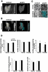
Figure 6. Disrupted remodeling of Purkinje dendrites in harmaline-treated mice.
Cellular morphology of P30 Purkinje cells in mice treated with saline (A) and harmaline (B) at P9–P14. Dendrites become irregular and multiplanar by harmaline treatment compared to the control littermate. Dendrites in minor sagittal planes are pseudocolored in graphic images. C: High power sagittal views of dendrites in respective boxed regions in A and B. Dendrites significantly overpass one another in harmaline-treated mice. D: Quantitative comparison of dendrite morphology in mice treated with saline or harmaline during P9–P14 or P15–P20. Overpassing branches are significantly increased by harmaline treatment in either period. Cells in the bank region of lobules IV and V were analyzed. n = 10 for each data point, mean±s.e.m, Student's t test, *p<0.01. Scale bars: 20 µm.

References

    1. Mainen ZF, Sejnowski TJ. Influence of dendritic structure on firing pattern in model neocortical neurons. Nature. 1996;382:363–366. - PubMed
    1. Stuart G, Spruston N, Hausser M. Oxford: Oxford University Press; 2007. Dendrites.
    1. Wei DS, Mei YA, Bagal A, Kao JP, Thompson SM, et al. Compartmentalized and binary behavior of terminal dendrites in hippocampal pyramidal neurons. Science. 2001;293:2272–2275. - PubMed
    1. Kaufmann WE, Moser HW. Dendritic anomalies in disorders associated with mental retardation. Cereb Cortex. 2000;10:981–991. - PubMed
    1. Parrish JZ, Emoto K, Kim MD, Jan YN. Mechanisms that regulate establishment, maintenance, and remodeling of dendritic fields. Annu Rev Neurosci. 2007;30:399–423. - PubMed

Publication types