. 2011;6(5):e20153.
doi: 10.1371/journal.pone.0020153. Epub 2011 May 31.

Chronic apocynin treatment attenuates beta amyloid plaque size and microglial number in hAPP(751)(SL) mice

Affiliations

Chronic apocynin treatment attenuates beta amyloid plaque size and microglial number in hAPP(751)(SL) mice

Melinda E Lull et al. PLoS One. 2011.

Abstract

Background: NADPH oxidase is implicated in neurotoxic microglial activation and the progressive nature of Alzheimer's Disease (AD). Here, we test the ability of two NADPH oxidase inhibitors, apocynin and dextromethorphan (DM), to reduce learning deficits and neuropathology in transgenic mice overexpressing human amyloid precursor protein with the Swedish and London mutations (hAPP(751)(SL)).

Methods: Four month old hAPP(751)(SL) mice were treated daily with saline, 15 mg/kg DM, 7.5 mg/kg DM, or 10 mg/kg apocynin by gavage for four months.

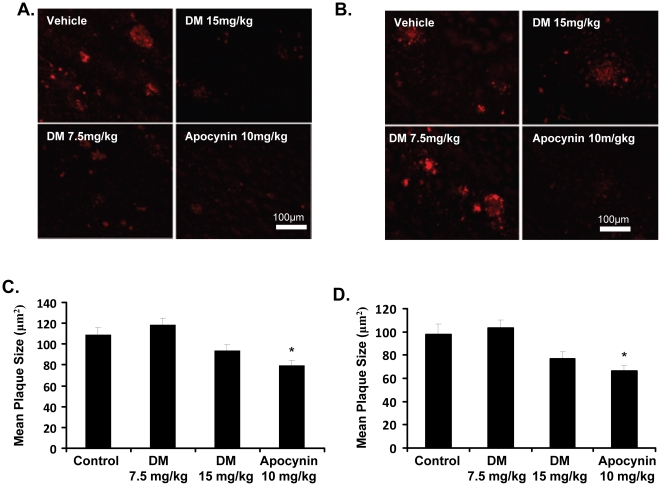
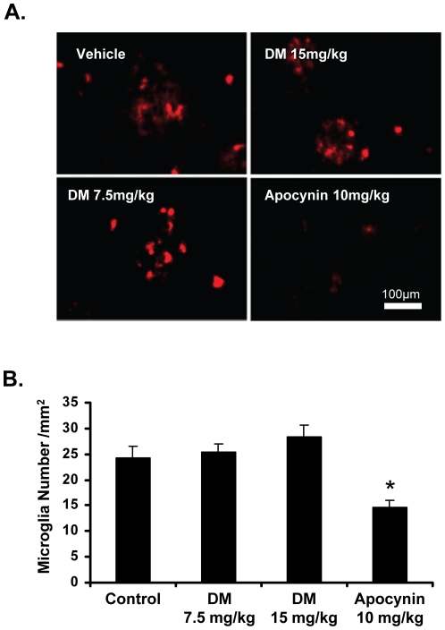
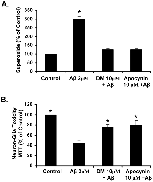
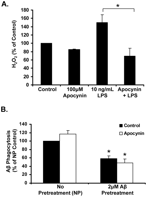
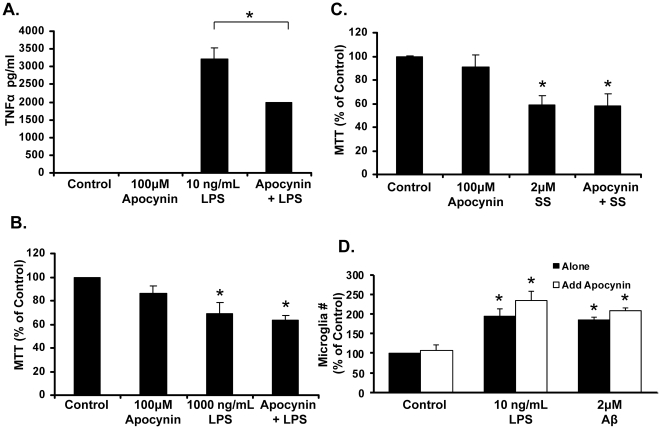
Results: Only hAPP(751)(SL) mice treated with apocynin showed reduced plaque size and a reduction in the number of cortical microglia, when compared to the saline treated group. Analysis of whole brain homogenates from all treatments tested (saline, DM, and apocynin) demonstrated low levels of TNFα, protein nitration, lipid peroxidation, and NADPH oxidase activation, indicating a low level of neuroinflammation and oxidative stress in hAPP(751)(SL) mice at 8 months of age that was not significantly affected by any drug treatment. Despite in vitro analyses demonstrating that apocynin and DM ameliorate Aβ-induced extracellular superoxide production and neurotoxicity, both DM and apocynin failed to significantly affect learning and memory tasks or synaptic density in hAPP(751)(SL) mice. To discern how apocynin was affecting plaque levels (plaque load) and microglial number in vivo, in vitro analysis of microglia was performed, revealing no apocynin effects on beta-amyloid (Aβ) phagocytosis, microglial proliferation, or microglial survival.

Conclusions: Together, this study suggests that while hAPP(751)(SL) mice show increases in microglial number and plaque load, they fail to exhibit elevated markers of neuroinflammation consistent with AD at 8 months of age, which may be a limitation of this animal model. Despite absence of clear neuroinflammation, apocynin was still able to reduce both plaque size and microglial number, suggesting that apocynin may have additional therapeutic effects independent of anti-inflammatory characteristics.


Conflict of interest statement

Competing Interests: The authors have declared that no competing interests exist.

Figures

Figure 1
Figure 1. Apocynin reduces plaque size in the cortex and hippocampus of hAPP(751)SL mice.
Mice were treated daily with 15 mg/kg dextromethorphan (DM), 7.5 mg/kg DM, or 10 mg/kg apocynin for four months. The size of β-amyloid plaques was measured for each group and compared to control, vehicle-treated animals. Representative images show 6E10 staining of β-amyloid protein for each group in the cortex (A) and hippocampus (B), respectively. Quantification of plaque size the cortex (C) and hippocampus (D) revealed that only apocynin significantly decreased the size of plaques, compared to vehicle. DM, at either dose, did not alter plaque size in the cortex or the hippocampus. Plaque size was determined as the absolute plaque area divided by the absolute plaque number. *p<0.05 vs. vehicle, 1-way ANOVA with Bonferroni post-hoc test.
Figure 2
Figure 2. Apocynin reduces the number of microglia in the cortex of hAPP(751)SL mice.
Mice were treated daily with 15 mg/kg dextromethorphan (DM), 7.5 mg/kg DM, or 10 mg/kg apocynin for four months. The number of microglia was then counted for each group by staining with anti-CD11b antibody and each treatment group was compared to control. CD11b-stained microglia were only counted if they corresponded to a DAPI stained nuclei (data not shown). Representative images from each group of the stained microglia are shown in panel (A). Apocynin reduced the number of microglia in the cortex of hAPP(751)SL mice, whereas neither dose of DM reduced microglia number (B). *p<0.05 vs vehicle, 1-way ANOVA with Bonferroni post-hoc test.
Figure 3
Figure 3. Apocynin reduces NADPH oxidase activation and is neuroprotective in vitro.
(A) Enriched microglia cultures were treated with media alone (Control), apocynin (10 µM), Dextromethorphan (DM, 10 µM), Aβ (2 µM), Apocynin + Aβ, and DM + Aβ. The production of extracellular superoxide was measured by the superoxide dismutase (SOD)-inhibitable reduction of tetrazolium salt, WST-1 at 30 minutes post-treatment. Results are mean ± SEM. Data are from four separate experiments. *p<0.05, compared with control cultures. (B) Apocynin and DM protect against Aβ-induced toxicity in cortical neuron-glia cultures.) Cortical neuron-glia cultures were treated with media alone (Control), Apocynin (10 µM), Dextromethorphan (DM, 10 µM), Aβ (2 µM), Apocynin + Aβ, and DM + Aβ. Toxicity was assessed by MTT 7 days later. Graphs show the results expressed as percentage of the control cultures and are the mean ± SEM from three independent experiments in triplicate. * p<0.05, control compared to treatment.
Figure 4
Figure 4. Apocynin regulates microglial H2O2 production, but not Aβ phagocytosis.
(A) Apocynin attenuates LPS-induced hydrogen peroxide (H2O2), as predicted. Microglia-enriched cultures were treated with Hank's balanced salt solution (HBSS), or HBSS with LPS (10 ng/mL), apocynin (100 µM), or the combination of apocynin (100 µM) and LPS for 3 hours. The level of H2O2 was then measured in each group and compared to control levels. Apocynin does significantly reduce LPS-induced increases in H2O2, returning levels to control values. *p<0.05 vs. control; #p<0.05 vs. LPS, 1-way ANOVA with Bonferroni post-hoc test. (B) Pre-treatment with 2 µM Aβ significantly reduces phagocytosis of fluorescent Aβ, and apocynin does not act to reverse this decrease. Microglia-enriched cultures were treated with control media, or media with β-amyloid (Aβ; 2 µM), apocynin (100 µM), or the combination of apocynin (100 µM) and Aβ (2 µM) for 24 hours. Fluorescently labeled Aβ (final concentration 0.1 µM) was then added to each well, and incubated with the cells for 6 hours to allow for phagocytosis of the fluorescent protein. The amount of phagocytosis of fluorescent Aβ was measured for each group and compared to control levels. *p<0.05 vs control, 1-way ANOVA with Bonferroni post-hoc test.
Figure 5
Figure 5. Apocynin ameliorates LPS-induced TNFα production, but has no effect on microglial cell death or cell number in vitro.
(A) Microglia-enriched cultures were treated with control media, lip polysaccharide (LPS; 10 ng/mL), and/or Apocynin (100 µM) for 24 hours. Tumor necrosis factor alpha (TNFα) levels in the supernatant were measured via ELISA. LPS (10 ng/mL) significantly increased levels of TNFα and pre-treatment with 100 µM Apocynin significantly reduced the amount of TNFα released by microglia. *p<0.05 vs. control; #p<0.05 vs. LPS, 1-way ANOVA with Bonferroni post-hoc test. (B) Apocynin does not protect against inflammation-induced cell death. Microglia-enriched cultures were treated with control media, 1000 ng/mL LPS, 100 µM Apocynin, or LPS and apocynin for 24 hours. After incubation, cell survival was measured with the MTT assay. 100 ng/mL LPS significantly reduced microglial cell survival (through inflammation-induced cell death), which is not rescued by apocynin). (C) Microglia-enriched cultures were treated with control media, 2 µM staurosporine (SS), 100 µM Apocynin, or SS and apocynin for 24 hours. After incubation, cell survival was measured with the MTT assay. Data show that 2 µM SS significantly reduces microglial cell survival (through apoptosis), and is not reversed by the addition of apocynin. *p<0.05 vs control,1-way ANOVA with Bonferroni post-hoc test. (D) Apocynin does not alter Aβ or LPS-induced increases in microglia number in vitro. Mixed neuron-glia cultures were treated with 2 µM Aβ, 10 ng/mL LPS and/or 100 µM apocynin for 24 hours. Cultures were then fixed and stained with IBA-1 antibody for microglia. The number of microglia was then counted in 9 representative areas per well. The number of microglia was significantly increased in cultures treated with LPS (196% of control) and Aβ (186% of control). Apocynin treatment did not prevent the increase in cell count caused by LPS or Aβ. Apocynin alone caused no significant change in cell count. *p<0.05 vs control, 1-way ANOVA with Bonferroni post-hoc test.

References

    1. Block ML. NADPH oxidase as a therapeutic target in Alzheimer's disease. BMC Neurosci. 2008;9(Suppl 2):S8. - PMC - PubMed
    1. Imbimbo BP, Solfrizzi V, Panza F. Are NSAIDs useful to treat Alzheimer's disease or mild cognitive impairment? Front Aging Neurosci. 2010;2 - PMC - PubMed
    1. Babior BM. Phagocytes and oxidative stress. Am J Med. 2000;109:33–44. - PubMed
    1. Qin L, Liu Y, Wang T, Wei SJ, Block ML, et al. NADPH oxidase mediates lipopolysaccharide-induced neurotoxicity and proinflammatory gene expression in activated microglia. J Biol Chem. 2004;279:1415–1421. - PubMed
    1. Wilkinson BL, Cramer PE, Varvel NH, Reed-Geaghan E, Jiang Q, et al. Ibuprofen attenuates oxidative damage through NOX2 inhibition in Alzheimer's disease. Neurobiol Aging. 2010. - PMC - PubMed

Publication types

MeSH terms