. 2011 May;9(5):e1001073.
doi: 10.1371/journal.pbio.1001073. Epub 2011 May 31.

N-terminal acetylation inhibits protein targeting to the endoplasmic reticulum

Affiliations

N-terminal acetylation inhibits protein targeting to the endoplasmic reticulum

Gabriella M A Forte et al. PLoS Biol. 2011 May.

Abstract

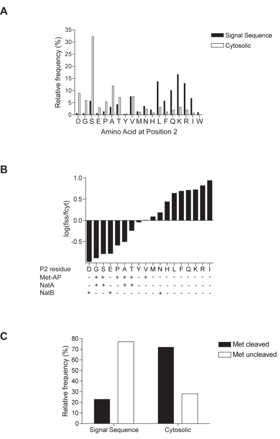
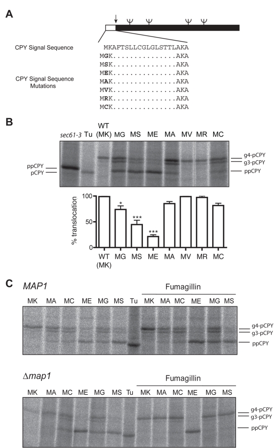
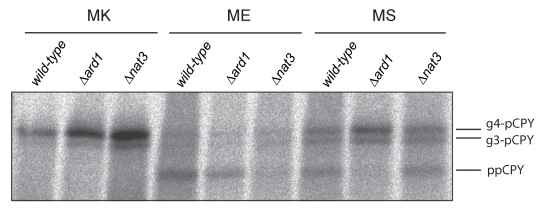
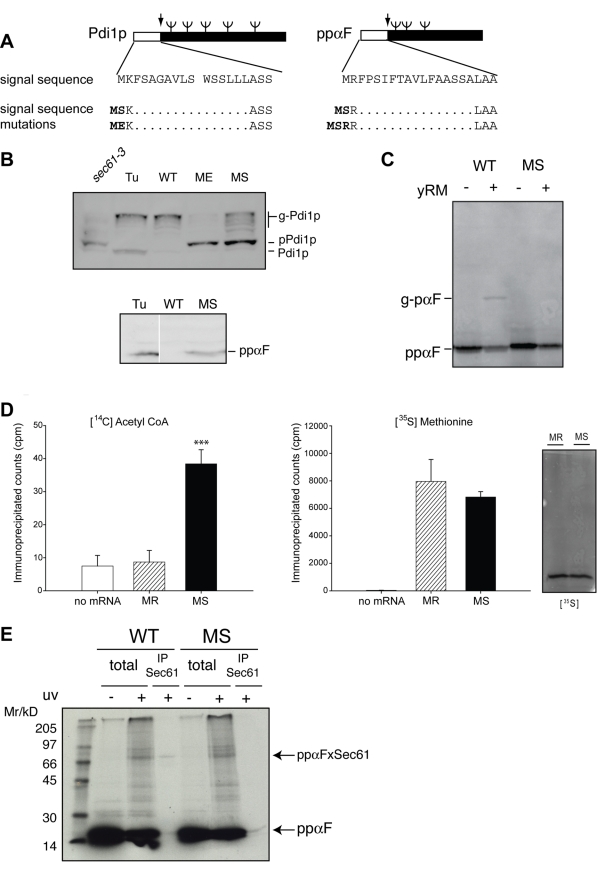
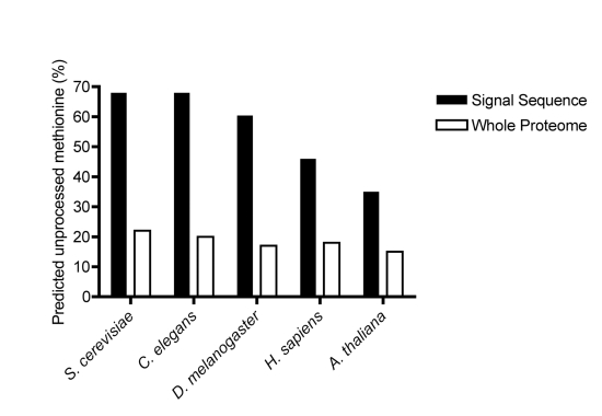
Amino-terminal acetylation is probably the most common protein modification in eukaryotes with as many as 50%-80% of proteins reportedly altered in this way. Here we report a systematic analysis of the predicted N-terminal processing of cytosolic proteins versus those destined to be sorted to the secretory pathway. While cytosolic proteins were profoundly biased in favour of processing, we found an equal and opposite bias against such modification for secretory proteins. Mutations in secretory signal sequences that led to their acetylation resulted in mis-sorting to the cytosol in a manner that was dependent upon the N-terminal processing machinery. Hence N-terminal acetylation represents an early determining step in the cellular sorting of nascent polypeptides that appears to be conserved across a wide range of species.


Conflict of interest statement

The authors have declared that no competing interests exist.

Figures

Figure 1
Figure 1. Amino acid frequency at P2 of signal sequences versus cytosolic proteins.
(A) Relative frequency of amino acids at P2 of a filtered set of 277 signal sequence-containing proteins from S. cerevisiae was compared to a similar size group (n = 252) of randomly selected cytosolic proteins. Frequency distribution between the groups differed significantly (p<0.0001, χ2 = 207.3 18 df). (B) Ratio of relative frequency of P2 residues between signal sequence (fss) and cytosolic (fcyt) proteins. Tryptophan was absent from the cytosolic group; therefore, no log(f ss/f cyt) value is plotted. P2 specificities of MetAP, NatA, and NatB are indicated. (C) Predicted methionine cleavage of signal sequence and cytosolic N-termini based on relative P2 frequency. For complete datasets, see Tables S1–S4.
Figure 2
Figure 2. Removal of the N-terminal methionine inhibits ER translocation of CPY.
(A) Schematic of wild-type CPY and P2 mutants. Signal peptide sequence, position of N-glycosylation (ψ), and signal peptidase cleavage (↓) sites are indicated. (B) Yeast cells (Δpep4,Δprc1) expressing either wild-type or mutant CPY were pulse-labelled with [35S]methionine/cysteine, then CPY immunoprecipitated, and analysed by SDS-PAGE and phosphorimaging. Positions of glycosylated CPY (g4-pCPY and g3-pCPY) are indicated as are the untranslocated ppCPY and signal-sequence cleaved, non-glycosylated CPY (pCPY) observed in sec61-3 cells and in tunicamycin-treated wild-type cells (Tu), respectively. Translocation efficiency was determined by quantification of ppCPY and g3- and g4-pCPY from three independent experiments. Error bars represent standard error of the mean. Asterisks represent p<0.05 (*) and p<0.001 (***) according to the one-way analysis of variance with Tukey's multiple comparison test. (C) CPY translocation was analysed as in (B), in a wild-type (Δpep4,Δprc1) and isogenic Δmap1 strain in the presence and absence of the Map2 inhibitor fumagillin (for quantification, see Figure S1).
Figure 3
Figure 3. N-terminal acetylation blocks protein translocation.
Translocation of wild-type, MS, and ME mutants of CPY was examined z(as in Figure 2B) in wild-type and Δard1 and Δnat3 strains, which lack NatA and NatB activity, respectively. Data are representative of three independent experiments.
Figure 4
Figure 4. Protein N-acetylation inhibits ER translocation both in vivo and in vitro.
(A) Schematic of wild-type and P2 signal sequence mutants of Pdi1p and preproα-factor. Position of N-glycosylation (ψ) and signal peptidase cleavage (↓) sites are indicated. (B) Wild-type and indicated mutants of myc-tagged Pdi1p and ppαF were expressed in wild-type (Δpep4) or sec61-3 strains, and treated, where indicated, with Tunicamycin (Tu). Steady-state levels of protein were determined by preparation of cell extracts from these strains and analysis by Western blot with anti-myc antibodies. (C) Wild-type (MR) and MS forms of lysine-less ppαF (where all lysines had been mutated to arginine) were translated in vitro, then incubated with yeast microsomes (yRM). Position of non-translocated (ppαF) and signal-sequence cleaved, glycosylated (g-pαF) are indicated. (D) Lysine-less forms of both wild-type (MR) and MS ppαF were translated in vitro in the presence of either [35S] methionine or [14C] acetyl-CoA and immuno-precipitated with anti-ppαF antibodies before analysis by either scintillation counting or SDS-PAGE. Error bars represent standard deviation; three asterisks indicate p<0.001 according to the two-tailed student's t test. (E) Wild-type (MR) and MS ppαF with lysine residues at positions 5 and 12 were translated in vitro in the presence of [35S] methionine and TDBA-lysyl-tRNA. Targeting to microsomes was performed in the absence of ATP and then cross-linking induced by uv-irradiation. Where indicated, samples were denatured and immuno-precipitated with Sec61 antisera.
Figure 5
Figure 5. An SRP-dependent precursor is refractory to N-acetylation.
(A) Schematic of wild-type OPY (CPY with the endogenous signal sequence replaced by that of Ost1) and corresponding P2 signal sequence mutants. (B) Wild-type and mutant OPY translocation in vivo was monitored by pulse-labelling and immunoprecipitation as in Figure 2B. (C) Lysine-less wild-type (MR) and MS opαF (ppαF with the signal sequence replaced with that of Ost1p and all lysines mutated to arginine) were translated in vitro in the presence of [35S] methionine, denatured, and modified with amine-reactive sulfo-NHS-SS-biotin. Biotinylated proteins were re-isolated on immobilized-streptavidin and analysed by SDS-PAGE and phosphorimaging.
Figure 6
Figure 6. A bias against N-terminal processing of signal sequences is conserved across eukaroytes.
Predicted frequency of an unprocessed initiating methionine in signal sequences from S. cerevisaie (n = 277), C. elegans (n = 378), Drosophila (n = 448), human (n = 595), and Arabidopsis (n = 500) compared to the respective proteomes as a whole . For complete datasets, see Tables S5 and S6.

References

    1. Giglione C, Boularot A, Meinnel T. Protein N-terminal methionine excision. Cell Mol Life Sci. 2004;61:1455–1474. - PMC - PubMed
    1. Martinez A, Traverso J. A, Valot B, Ferro M, Espagne C, et al. Extent of N-terminal modifications in cytosolic proteins from eukaryotes. Proteomics. 2008;8:2809–2831. - PubMed
    1. Arnesen T, Van Damme P, Polevoda B, Helsens K, Evjenth R, et al. Proteomics analyses reveal the evolutionary conservation and divergence of N-terminal acetyltransferases from yeast and humans. Proc Natl Acad Sci U S A. 2009;106:8157–8162. - PMC - PubMed
    1. Arfin S. M, Bradshaw R. A. Cotranslational processing and protein turnover in eukaryotic cells. Biochemistry. 1988;27:7979–7984. - PubMed
    1. Hwang C. S, Shemorry A, Varshavsky A. N-erminal acetylation of cellular proteins creates specific degradation signals. Science. 2010;327:973–977. - PMC - PubMed

Publication types

MeSH terms

Substances