. 2011 Dec;7(4):427-34.
doi: 10.1007/s11302-011-9239-6. Epub 2011 Jun 9.

Liver damage and systemic inflammatory responses are exacerbated by the genetic deletion of CD39 in total hepatic ischemia

Affiliations

Liver damage and systemic inflammatory responses are exacerbated by the genetic deletion of CD39 in total hepatic ischemia

Xiaofeng Sun et al. Purinergic Signal. 2011 Dec.

Abstract

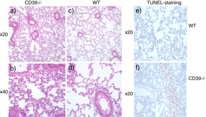
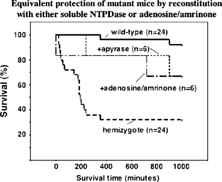
Liver ischemia reperfusion injury is associated with both local damage to the hepatic vasculature and systemic inflammatory responses. CD39 is the dominant vascular endothelial cell ectonucleotidase and rapidly hydrolyses both adenosine triphosphate (ATP) and adenosine diphosphate to adenosine monophosphate. These biochemical properties, in tandem with 5'-nucleotidases, generate adenosine and potentially illicit inflammatory vascular responses and thrombosis. We have evaluated the role of CD39 in total hepatic ischemia reperfusion injury (IRI). Wildtype mice, Cd39-hemizygous mice (+/-) and matched Cd39-null mice (-/-); (n = 25 per group) underwent 45 min of total warm ischemia with full inflow occlusion necessitating partial hepatectomy. Soluble nucleoside triphosphate diphosphohydrolase (NTPDases) or adenosine/amrinone were administered to wildtype (n = 6) and Cd39-null mice (n = 6) in order to study protective effects in vivo. Parameters of liver injury, systemic inflammation, hepatic ATP determinations by P(31)-NMR and parameters of lung injury were obtained. All wildtype mice survived up to 7 days with minimal biochemical disturbances and minor evidence for injury. In contrast, 64% of Cd39+/- and 84% of Cd39-null mice required euthanasia or died within 4 h post-reperfusion with liver damage and systemic inflammation associated with hypercytokinemia. Hepatic ATP depletion was pronounced in Cd39-null mice posthepatic IRI. Soluble NTPDase or adenosine administration protected Cd39-deficient mice from acute reperfusion injury. We conclude that CD39 is protective in hepatic IRI preventing local injury and systemic inflammation in an adenosine dependent manner. Our data indicate that vascular CD39 expression has an essential protective role in hepatic IRI.


Figures

Fig. 1
Fig. 1
Cd39-deficient (P < 0.001) and null (P < 0.05) mice exhibit significantly higher rates of mortality after 45 min of warm full hepatic inflow ischemia and consecutive reperfusion than the matched wildtype mice
Fig. 2
Fig. 2
a Wild-type livers after 45 min of ischemia and 4 h reperfusion show patchy macroscopic hemorrhage. In contrast, the Cd39-deficient livers show overt areas of infarcted parenchyma from hepatic venous thrombosis (b). a Sinusoidal congestion is seen in the wildtype mouse post hepatic ischemia and reperfusion (IRI) (H&E staining). b The CD39-null mice show massive sinusoidal hemorrhagic necrosis post IRI (H&E staining). c Fibrin staining of non-infarcted hepatic parenchyma using T2G1 immunohistochemical staining showing patchy areas of fibrin deposition. d Abundant fibrin deposition in the hepatic parenchyma in the Cd39-deficient mice, suggesting hepatic parenchymal cell injury. TUNEL staining for apoptosis demonstrates minimal apoptosis in the wild-type animals (e), but large apoptotic areas in the CD39−/− mice (f)
Fig. 3
Fig. 3
H&E staining of lung tissue collected after 45 min of full hepatic ischemia and 4 h of reperfusion. It is clearly visible that the Cd39-null animals (a, b) show increased alveolar edema compared with the wildtype controls (c, d). TUNEL-staining indicative of apoptosis demonstrates areas of apoptosis in the Cd39-null (f) lung tissue after 45 min of full hepatic ischemia and 4 h of reperfusion, when compared to the wildtype control (e)
Fig. 4
Fig. 4
Systemic inflammation characterized by elevated pro inflammatory cytokines is noted in Cd39-hemizygous and null mice after 45 min of hepatic ischemia and 4 h of reperfusion. IL-6 (p < 0.02) and TNF (p < 0.01) are significantly increased in Cd39-null mice compared to the wildtype controls. Significant increases in IL-1 in the Cd39-hemizygous (p < 0.007) and Cd39-null mice (p < 0.005) compared to wildtype animals are evident
Fig. 5
Fig. 5
Hepatic reperfusion injury: NMR – P31 spectra with ATP depletion in the wild-type mouse. 31P spectra from the left lateral mouse liver lobe at the three different time frames (top) pre-ischemia, (middle) 45 min of ischemia, and (bottom) 30–60 min of reperfusion. The intensity of the three peaks due to the abg ATP is reduced during ischemia and recovery in the wild-type upon reperfusion. In contrast, the recovery of this metabolite in the Cd39-null mouse liver appears to be absent or comparatively slow. Furthermore, the increase in inorganic phosphate signal (Pi) due to anoxia/hypoxia during the ischemic episode in both types of mice, which recover in the wild type and remain elevated in the heterozygous even after 12reperfusion
Fig. 6
Fig. 6
The administration of apyrase improves survival in Cd39-hemizygous mice when injected intravenously pre-reperfusion. Survival of Cd39-null animals could not be dramatically improved after apyrase injection (data not shown). The administration of adenosine also markedly improved Cd39-hemizygous survival after hepatic IRI
Fig. 7
Fig. 7
AST levels after 45 min of hepatic ischemia and reperfusion. The Cd39-deficient mice show a significant increase of AST levels, when compared to wildtype mice at 15 min (p < 0.0005) and at 1 h of reperfusion (p < 0.0003). After administration of apyrase AST levels are significantly decreased in the Cd39-deficient group when compared to the Cd39-deficient group at 15 min (p < 0.003) and at 1 h of reperfusion (p < 0.0004)

References

    1. Thiagarajan RR, Winn RK, Harlan JM. The role of leukocyte and endothelial adhesion molecules in ischemia-reperfusion injury. Thromb Haemost. 1997;78:310–314. - PubMed
    1. Milazzo VJ, Ferrante RJ, Sabido F, et al. Time course of leukocyte adhesion to endothelium in ischemia-reperfusion. J Surg Res. 1996;61:139–142. doi: 10.1006/jsre.1996.0094. - DOI - PubMed
    1. Squadrito F, Altavilla D, Squadrito G, et al. The involvement of tumour necrosis factor-alpha in the protective effects of 17 beta oestradiol in splanchnic ischaemia-reperfusion injury. Br J Pharmacol. 1997;121:1782–1788. doi: 10.1038/sj.bjp.0701288. - DOI - PMC - PubMed
    1. Rose S, Floyd RA, Eneff K, et al. Intestinal ischemia: reperfusion-mediated increase in hydroxyl free radical formation as reported by salicylate hydroxylation. Shock. 1994;1:452–456. doi: 10.1097/00024382-199406000-00010. - DOI - PubMed
    1. Kaczmarek E, Erb L, Koziak K, et al. Modulation of endothelial cell migration by extracellular nucleotides: involvement of focal adhesion kinase and phosphatidylinositol 3-kinase-mediated pathways. Thromb Haemost. 2005;93:735–742. - PMC - PubMed