. 2011 Dec;66(6):1777-87.
doi: 10.1002/mrm.22961. Epub 2011 Jun 7.

Size-optimized 32-channel brain arrays for 3 T pediatric imaging

Affiliations

Size-optimized 32-channel brain arrays for 3 T pediatric imaging

Boris Keil et al. Magn Reson Med. 2011 Dec.

Abstract

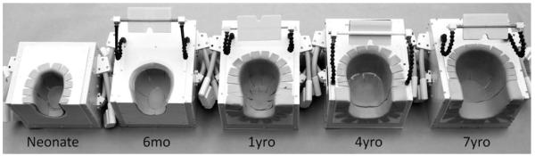
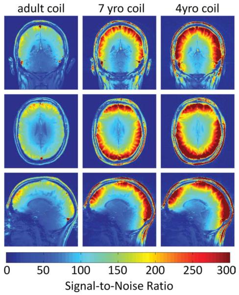
Size-optimized 32-channel receive array coils were developed for five age groups, neonates, 6 months old, 1 year old, 4 years old, and 7 years old, and evaluated for pediatric brain imaging. The array consisted of overlapping circular surface coils laid out on a close-fitting coil-former. The two-section coil former design was obtained from surface contours of aligned three-dimensional MRI scans of each age group. Signal-to-noise ratio and noise amplification for parallel imaging were evaluated and compared to two coils routinely used for pediatric brain imaging; a commercially available 32-channel adult head coil and a pediatric-sized birdcage coil. Phantom measurements using the neonate, 6-month-old, 1-year-old, 4-year-old, and 7-year-old coils showed signal-to-noise ratio increases at all locations within the brain over the comparison coils. Within the brain cortex the five dedicated pediatric arrays increased signal-to-noise ratio by up to 3.6-, 3.0-, 2.6-, 2.3-, and 1.7-fold, respectively, compared to the 32-channel adult coil, as well as improved G-factor maps for accelerated imaging. This study suggests that a size-tailored approach can provide significant sensitivity gains for accelerated and unaccelerated pediatric brain imaging.


Figures

FIG. 1
FIG. 1
The completed array coil for 1 year old consists of two segments; a deep posterior segmentand a frontal paddle over the forehead (in orange). The eyes and face of the subject are completely unobstructed. a: Finalized coil enclosed in a plastic box; b: inside view of the three-dimensional printed coil formers with coil circuitry; c: tiling geometry diagram of the 32-channel layout; the loop diameters are slightly larger than the diameter of the circle which inscribe the vertexes of the hexagon/pentagons. [Color figure can be viewed in the online issue, which is available at wileyonlinelibrary.com.]
FIG. 2
FIG. 2
Posterior coil segments of all five constructed pediatric array coils. The coil former arebased on the 95th percentile MRI contours of corresponding aged children and have been dilated to accommodate foam padding. The coil formers were three-dimensional printed and enclosed in a plastic box. Mounted mirrors are used to project visual stimulus for research studies.
FIG. 3
FIG. 3
Circuit schematic for the coil element and preamplifier chain. The coil element uses three capacitors: The variable capacitor C1 fine-tunes the coil element frequency. Where C2 and C3 are equally valued they provide a capacitive voltage divider at the coil output circuit. A detuning trap is formed around C2 using a variable inductor L and a Diode D. A small coaxial cable connects coil and preamp transforming the element impedance to ZNM, the noise matched impedance for the preamplifier input. The coaxial cable also transforms the input impedance of the preamplifier to a short across D.
FIG. 4
FIG. 4
Noise correlation matrices of all five constructed pediatric 32-channel brain arrays acquired with the size-matched loading phantoms. The overall average noise correlation (all coils) is 12%. [Color figure can be viewed in the online issue, which is available at wileyonlinelibrary.com.]
FIG. 5
FIG. 5
SNR comparisons between sagittal images obtained from the sized-matched head phantoms using the pediatric brain array coils (first row), the 32-channel adult brain array coil (second row), and a CP birdcage coil (third row). The images show that the highest SNR gain occurs closest to the surface of the constructed array. In the “brain” center of the phantoms the SNR is only slightly improved. The superimposed ROI on the birdcage coil SNR maps correspond to the regions used in the average brain SNR measurement.The 7-year-old phantom was too big to fit into the pediatric birdcage coil. [Color figure can be viewed in the online issue, which is available at wileyonlinelibrary.com.]
FIG. 6
FIG. 6
In vivo SNR comparisons in sagittal, axial, and coronal planes between a commercial available 32-channel adult coil, the 7-year-old coil, and the 4-year-old coil using a same small head-sized adult in all coils. Compared to pediatric heads, the adult head is slightly bigger in anterior–posterior direction. This leads to a more prominent gap between frontal paddle elements and the posterior elements. The in vivo measurements match the phantom SNR results and verify that a sized matched array approach shows a peripheral SNR increase. [Color figure can be viewed in the online issue, which is available at wileyonlinelibrary.com.]
FIG. 7
FIG. 7
Transverse maps of 1/G-factor obtained from the constructed array coils and the adult coil using the age-matched head phantoms. The FOVs were chosen as tight as possible during image acquisition to avoid underestimation of G-factors. The maps were calculated using images from PD-weighted GRE sequence and noise correlation information. The pediatric brain arrays show over-all lower average and peak G-factor values compared with the 32-channel adult coil.
FIG. 8
FIG. 8
Average and peak G-factors for adult and pediatric 32-channel arrays from the data in FIG. 7. a: Whole-slice average G-factors comparing the overall noise amplification during parallel image reconstruction. b: Peak G-factors for the slice, reflecting the worst-case noise amplification. c, d: G-factor averages in a central and peripheral ROI. The highest relative gain by going to the size-appropriate coil was achieved in the central brain region. The pediatric coils show overall more favorable peak G-factors for both one-dimensional and two-dimensional accelerations directions. The scans were accelerated in the anterior–posterior direction (for one-dimensional acceleration) and anterior–posterior and right-left direction for the two-dimensional accelerations. [Color figure can be viewed in the online issue, which is available at wileyonlinelibrary.com.]
FIG. 9
FIG. 9
Fully encoded sagittal 1-mm isotropic MPRAGE images with GRAPPA acceleration factor of R = 3. For coil performance comparison, the images were acquired with the 4-year-old (a, c) and the adult 32-channel (b, d) array coils using the same adult subject. Corresponding close ups (c, d) show better image details using the size matched array coil for 4-year-old children.
FIG. 10
FIG. 10
T2-weighted transverse high resolution (0.4 × 0.4 × 2 mm3) turbo spin echo (TSE) images, acquired using the neonate and 4-year-old coils. Both images have been intensity normalized. a: Four-day old sedated neonate imaged in 6:58 min using the 32-channel neonate array for a separate clinical study. b: Image acquired (4:16 min) in the 4-year-old array of an adult with a small enough head to fit this coil.

References

    1. Roemer PB, Edelstein WA, Hayes CE, Souza SP, Mueller OM. The NMR phased array. Magn Reson Med. 1990;16:192–225. - PubMed
    1. Hayes CE, Hattes N, Roemer PB. Volume imaging with MR phased arrays. Magn Reson Med. 1991;18:309–319. - PubMed
    1. Wald LL, Carvajal L, Moyher SE, Nelson SJ, Grant PE, Barkovich AJ, Vigneron DB. Phased array detectors and an automated intensity-correction algorithm for high-resolution MR imaging of the human brain. Magn Reson Med. 1995;34:433–439. - PubMed
    1. Griswold MA, Jakob PM, Heidemann RM, Nittka M, Jellus V, Wang J, Kiefer B, Haase A. Generalized autocalibrating partially parallel acquisitions (GRAPPA) Magn Reson Med. 2002;47:1202–1210. - PubMed
    1. Pruessmann KP, Weiger M, Scheidegger MB, Boesiger P. SENSE: sensitivity encoding for fast MRI. Magn Reson Med. 1999;42:952–962. - PubMed

Publication types