Combined genomic and phenotype screening reveals secretory factor SPINK1 as an invasion and survival factor associated with patient prognosis in breast cancer
- PMID: 21656687
- PMCID: PMC3377086
- DOI: 10.1002/emmm.201100150
Combined genomic and phenotype screening reveals secretory factor SPINK1 as an invasion and survival factor associated with patient prognosis in breast cancer
Abstract
Secretory factors that drive cancer progression are attractive immunotherapeutic targets. We used a whole-genome data-mining approach on multiple cohorts of breast tumours annotated for clinical outcomes to discover such factors. We identified Serine protease inhibitor Kazal-type 1 (SPINK1) to be associated with poor survival in estrogen receptor-positive (ER+) cases. Immunohistochemistry showed that SPINK1 was absent in normal breast, present in early and advanced tumours, and its expression correlated with poor survival in ER+ tumours. In ER- cases, the prognostic effect did not reach statistical significance. Forced expression and/or exposure to recombinant SPINK1 induced invasiveness without affecting cell proliferation. However, down-regulation of SPINK1 resulted in cell death. Further, SPINK1 overexpressing cells were resistant to drug-induced apoptosis due to reduced caspase-3 levels and high expression of Bcl2 and phospho-Bcl2 proteins. Intriguingly, these anti-apoptotic effects of SPINK1 were abrogated by mutations of its protease inhibition domain. Thus, SPINK1 affects multiple aggressive properties in breast cancer: survival, invasiveness and chemoresistance. Because SPINK1 effects are abrogated by neutralizing antibodies, we suggest that SPINK1 is a viable potential therapeutic target in breast cancer.
Copyright © 2011 EMBO Molecular Medicine.
Figures

SPINK1 expression in all samples.
ER-negative cases.
ER-positive cases.

SPINK1 expression was negligible in 10 normal breast cores (panels a–h).
This panel shows representative cores from the commercial breast TMA. As shown, invasive ductal carcinomas were positive but displayed various levels of SPINK1 (panels a–h).
SPINK1 nuclear staining was largely restricted to the breast tumour cells (green arrow) as compared to adjacent normal cells (red arrow).
Localization of SPINK1 in vitro. MDA-MB-231 cells were treated with SPINK1-CM and uptake of SPINK1 (if any) was studied using two antibodies. SPINK1 staining observed in cells after 12–24 h of SPINK1 treatment, recapitulating staining observed in primary tumours (Blue: DAPI; Green: anti-SPINK1 antibody; Red: anti-His antibody).

SPINK1 protein expression in all samples.
ER-negative cases.
ER-positive cases.

siRNA-mediated knockdown (kd) of SPINK1 using three different siRNAs in breast cancer cell lines MCF-7 and MDA-MB-231. SPINK1 expression was detected via RT-PCR.
Effect of SPINK1-kd on cell proliferation in MDA-MB-231 (left panel) and MCF-7 (right panel).
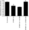
Activated PARP and caspase-3 upon siRNA-mediated knockdown of SPINK1 in MDA-MB-231 (left panel) and MCF-7 (right panel). (*p < 0.005; **p < 0.05; ***p < 1E−05).
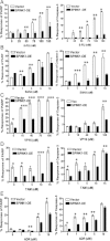
5-FU (5-fluorouracil)
SAHA (suberoylanilide hydoramic acid)
VP16 (etoposide)
TAM (tamoxifen)
ADR (adriamycin)

Protein levels of main apoptotic regulators were measured via Western blot 2 h after SPINK1CM (SH9), vecCM or no CM was added to MCF-7 cells.
Expression of main apoptotic regulators was measured in MCF-7 cells that overexpress SPINK1 at varying levels (SH5 and SH9), along with empty vector overexpression as a control.

5-FU
SAHA
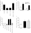
Cell invasion assay with MDA-MB-231 cells. Loss of invasion upon SPINK1 siRNA treatment, and rescued with SPINK1-CM.
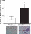
Effect of SPINK1-CM on invasion in two breast cell lines. The total number of cells that invaded was measured after 24 h (MB231) or 48 h (BT549) in response to SPINK1-CM and vector control CM.
Effect of SF9spink1CM and immunoneutralization on MDA-MB-231 cells. (White bars—SF9vecCM; black bars—SF9spink1CM diluted 10×; checkered bars—CM with addition of neutralizing antibody against SPINK1).
Invasion of MDA-MB-231 treated with WT SPINK1 and mutant SPINK1. Number of cells invaded were measured 24 h after assay setup. (*p < 0.005; **p < 0.05).
References
-
- Bagul A, Pushpakom S, Balachander S, Newman WG, Siriwardena A. The SPINK1 N34S variant is associated with acute pancreatitis. Eur J Gastroenterol Hepatol. 2009;21:485. - PubMed
-
- Chin K, DeVries S, Fridlyand J, Spellman PT, Roydasgupta R, Kuo WL, Lapuk A, Neve RM, Qian Z, Ryder T, et al. Genomic and transcriptional aberrations linked to breast cancer pathophysiologies. Cancer Cell. 2006;10:529–541. - PubMed
-
- Das K, Mohd Omar MF, Ong CW, Bin Abdul Rashid S, Peh BK, Putti TC, Tan PH, Chia KS, Teh M, Shah N, et al. TRARESA: a tissue microarray-based hospital system for biomarker validation and discovery. Pathology. 2008;40:441–449. - PubMed
Publication types
MeSH terms
Substances
LinkOut - more resources
Full Text Sources
Other Literature Sources
Medical
Molecular Biology Databases
Research Materials

