. 2011 Jun 9;70(5):855-62.
doi: 10.1016/j.neuron.2011.03.025.

Projection-specific modulation of dopamine neuron synapses by aversive and rewarding stimuli

Affiliations

Projection-specific modulation of dopamine neuron synapses by aversive and rewarding stimuli

Stephan Lammel et al. Neuron. .

Abstract

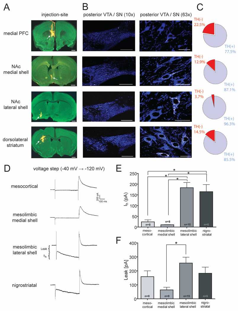
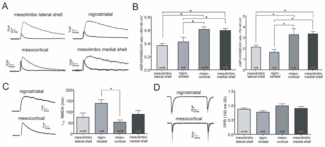
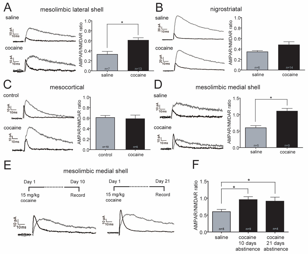
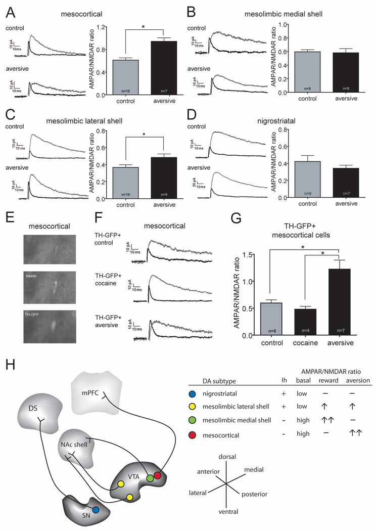
Midbrain dopamine (DA) neurons are not homogeneous but differ in their molecular properties and responses to external stimuli. We examined whether the modulation of excitatory synapses on DA neurons by rewarding or aversive stimuli depends on the brain area to which these DA neurons project. We identified DA neuron subpopulations in slices after injection of "Retrobeads" into single target areas of adult mice and found differences in basal synaptic properties. Administration of cocaine selectively modified excitatory synapses on DA cells projecting to nucleus accumbens (NAc) medial shell while an aversive stimulus selectively modified synapses on DA cells projecting to medial prefrontal cortex. In contrast, synapses on DA neurons projecting to NAc lateral shell were modified by both rewarding and aversive stimuli, which presumably reflects saliency. These results suggest that the mesocorticolimbic DA system may be comprised of three anatomically distinct circuits, each modified by distinct aspects of motivationally relevant stimuli.


Figures

Figure 1
Figure 1. Retrograde Labeling of Midbrain DA Neurons
(A) Injection sites (arrows) in green nissl (488 nm) counterstained sections (100 µm) showing typical locations of Retrobeads (546 nm, yellow). From top to bottom: mPFC (bregma +1.70 mm), NAc medial shell (bregma +1.10 mm), NAc lateral shell (bregma +0.74 mm), dorsolateral striatum (bregma +0.98 mm). Scale bars 500 µm. (B) Confocal images showing the anatomical distribution of retogradely transported Retrobeads (white) in the posterior VTA and SN following TH-immunohistochemistry (blue) at low magnification (10X, left panels) and high magnification (63X, right panels). Note that cells projecting to the mPFC are located in medial aspects of the posterior VTA and cells projecting to the NAc medial shell are located in the ventromedial areas of the posterior VTA. In contrast, neurons projecting to the NAc lateral shell are located in the dorsolateral region of the posterior VTA and cells projecting to the dorsolateral striatum are completely located in the posterior SN. Scale bars 200 µm (left panel) and 20 µm (right panel). (C) Pie charts showing the relative number of retrogradely labeled TH-immunopositive and TH-immunonegative cells that were located in the posterior VTA or posterior SN (Bregma −3.80 to −3.28 mm) (mesocortical n=49 cells; mesolimbic medial shell n=101 cells; mesolimbic lateral shell n=107 cells; nigrostriatal n=140 cells). (D) Representative current traces in response to a voltage step from −40 to −120 mV. Measurements of IRK+Leak currents and Ih are indicated in the mesolimbic lateral shell trace. (E) Magnitude of Ih for each cell population. Number of cells are indicated (* p<0.05). (F) Magnitude of IRK + leak currents for each cell population with number of cells indicated (* p<0.05).
Figure 2
Figure 2. Excitatory Synapses on DA Neurons Subpopulations Have Distinct Properties
(A) Sample AMPAR- and NMDAR EPSCs at +40 mV from different subpopulations of DA neurons. (B) AMPAR/NMDAR ratios at +40 mV (left panel) and at −70 mV/+40 mV (right panel) in the different DA neuron subpopulations. Number of cells are indicated (* p<0.05). (C) Sample NMDAR EPSCs at +40 mV for nigrostriatal and mesocortical DA neurons (left). Weighted decay time constants (τW) of NMDAR EPSCs recorded at +40 mV for all DA neuron subpopulations (right). Number of cells are indicated (*p<0.05). (D) Sample AMPAR EPSCs at −70 mV in response to paired afferent stimuli (100 ms interstimulus interval, ISI) for nigrostriatal and mesocortical DA neurons (left). Calculated paired-pulse ratios (PPR) at 100 ms ISI for all DA neuron subpopulations were not significantly different (right). Number of cells are indicated.
Figure 3
Figure 3. Cocaine Administration Increases AMPAR/NMDAR Ratios Only in DA Cells Projecting to NAc
(A–D) Sample AMPAR- and NMDAR EPSCs (left panels) and magnitude of AMPAR/NMDAR ratios (right panels) in different DA neuron subpopulations in animals that received saline or cocaine injections 24 hours prior to slice preparation. Number of cells are indicated (*p<0.05). Note that DA cells projecting to NAc lateral shell (A) and NAc medial shell (D) showed large increases in AMPAR/NMDAR ratios but nigrostriatal cells (B) and cells projecting to mPFC (C) did not. (The control cells in C are the same as those shown in Figure 2B.) (E) Sample AMPAR- and NMDAR EPSCs recorded from DA cells projecting to NAc medial shell 10 or 21 days after cocaine administration (F) Magnitude of AMPAR/NMDAR ratios at these time points compared to saline injected animals. Number of cells are indicated (*p<0.05).
Figure 4
Figure 4. An Aversive Stimulus Increases AMPAR/NMDAR Ratio in DA Cells Projecting to the mPFC
(A–D) Sample AMPAR- and NMDAR EPSCs (left panels) and magnitude of AMPAR/NMDAR ratios (right panels) in different DA neuron subpopulations in animals that received hindpaw formalin injections 24 hours prior to slice preparation. Number of cells are indicated (*p<0.05). Note that DA cells projecting to the mPFC (A) and NAc lateral shell (C) showed increases in AMPAR/NMDAR ratios but nigrostriatal cells (D) and cells projecting to NAc medial shell (B) did not. (The control cells in there panels are the same as those shown in Figure 2B.) (E) Sample GFP+ cell containing Retrobeads that were injected into the mPFC of a TH-GFP mouse 21 days earlier. Images obtained from a living brain slice using infrared videomicroscopy and epifluorescence. (F) Sample AMPAR- and NMDAR EPSCs recorded from GFP+ mesocortical cells in slices prepared from TH-GFP mice that had received cocaine or hindpaw formalin injections 24 hours earlier. (G) Magnitude of AMPAR/NMDAR ratios in GFP+ cells from control TH-GFP mice or mice that had received cocaine or the aversive formalin injections 24 hours earlier. Number of cells are indicated (*p<0.05). (H) Summary of some of the basal properties of the DA cell subpopulations and the modulation of their excitatory synapses by rewarding and aversive stimuli. Note that DA neurons projecting to the NAc lateral shell can be found in the anterior and posterior VTA (yellow dots), while DA neurons projecting to the mPFC and NAc medial shell are mainly located in the posterior VTA (red and green dots).

Comment in

References

    1. Abercrombie ED, Keefe KA, DiFrischia DS, Zigmond MJ. Differential effect of stress on in vivo dopamine release in striatum, nucleus accumbens, and medial frontal cortex. J. Neurochem. 1989;52:1655–1658. - PubMed
    1. Aragona BJ, Cleaveland NA, Stuber GD, Day JJ, Carelli RM, Wightman RM. Preferential enhancement of dopamine transmission within the nucleus accumbens shell by cocaine is attributable to a direct increase in phasic dopamine release events. J. Neurosci. 2008;28:8821–8831. - PMC - PubMed
    1. Argilli E, Sibley DR, Malenka RC, England PM, Bonci A. Mechanism and time course of cocaine-induced long-term potentiation in the ventral tegmental area. J. Neurosci. 2008;28:9092–9100. - PMC - PubMed
    1. Bassareo V, De Luca MA, Di Chiara G. Differential expression of motivational stimulus properties by dopamine in nucleus accumbens shell versus core and prefrontal cortex. J. Neurosci. 2002;22:4709–4719. - PMC - PubMed
    1. Bellone C, Luscher C. Cocaine triggered AMPA receptor redistribution is reversed in vivo by mGluR-dependent long-term depression. Nat. Neurosci. 2006;9:636–641. - PubMed

Publication types

MeSH terms