. 2011 Jun 9;70(5):966-78.
doi: 10.1016/j.neuron.2011.04.014.

VEGF mediates commissural axon chemoattraction through its receptor Flk1

Affiliations

VEGF mediates commissural axon chemoattraction through its receptor Flk1

Carmen Ruiz de Almodovar et al. Neuron. .

Erratum in

  • VEGF Mediates Commissural Axon Chemoattraction through Its Receptor Flk1.
    Ruiz de Almodovar C, Fabre PJ, Knevels E, Coulon C, Segura I, Haddick PCG, Aerts L, Delattin N, Strasser G, Oh WJ, Lange C, Vinckier S, Haigh J, Fouquet C, Gu C, Alitalo K, Castellani V, Tessier-Lavigne M, Chedotal A, Charron F, Carmeliet P. Ruiz de Almodovar C, et al. Neuron. 2023 Apr 19;111(8):1348. doi: 10.1016/j.neuron.2023.03.029. Neuron. 2023. PMID: 37080170 No abstract available.

Abstract

Growing axons are guided to their targets by attractive and repulsive cues. In the developing spinal cord, Netrin-1 and Shh guide commissural axons toward the midline. However, the combined inhibition of their activity in commissural axon turning assays does not completely abrogate turning toward floor plate tissue, suggesting that additional guidance cues are present. Here we show that the prototypic angiogenic factor VEGF is secreted by the floor plate and is a chemoattractant for commissural axons in vitro and in vivo. Inactivation of Vegf in the floor plate or of its receptor Flk1 in commissural neurons causes axon guidance defects, whereas Flk1 blockade inhibits turning of axons to VEGF in vitro. Similar to Shh and Netrin-1, VEGF-mediated commissural axon guidance requires the activity of Src family kinases. Our results identify VEGF and Flk1 as a novel ligand/receptor pair controlling commissural axon guidance.


Figures

Figure 1
Figure 1. VEGF is expressed at the floor plate
(A) In situ hybridization of VEGF mRNA on a E11.5 mouse spinal cord transverse section showing expression of VEGF at the floor plate (black arrow; also shown at higher magnification in inset), in the ventral spinal cord (red arrow) and in the motor columns (asterisks). (B) A sense probe, used as negative control, did not give any background signal. (C) β-Gal immunostaining (red) of a E11.5 VEGFLacZ mouse spinal cord transverse section at low and high (inset) magnification, revealing expression of β-Gal in the ventral spinal cord (yellow arrow), motor columns (asterisks) and floor plate (white arrow). (D) Bar graphs showing VEGF protein levels, released in floor plate conditioned medium (FPcm) from E11.5 wt mouse embryos, but not in control medium (Ctrl). Scale bars: A–C, 100 μm; insets A,C, 50 μm.
Figure 2
Figure 2. Inactivation of VEGF in the floor plate causes axon guidance defects in vivo
(AD) Robo3 immunostaining (red) in embryos with inactivation of one allele of Vegf in the floor plate (VegfFP-he; generated by crossing Hoxa1-Cre with Vegflox/lox mice) and in control embryos (VegfFP-wt). Compared to VegfFP-wt embryos (A,C), commissural axons in VegfFP-he embryos are defasciculated and some axons project near the lateral edge of the spinal cord (white arrows) (B,D). Panels C,D are higher magnification of the insets in A,B. (E) Scheme depicting the observed phenotypes. Left: normal commissural axons (red) project from the dorsal spinal cord to the floor plate (blue) in VegfFP-wt embryos. Right: in VegfFP-he embryos, axons project in a highly disorganized and defasciculated manner. (F) Left: histogram shows the quantification of the area occupied by commissural axons (% of total spinal cord area), normalized to the values obtained in VegfFP-wt embryos (see experimental procedures). *: P=0.03, Student’s t-test; N=8 VegfFP-wt and N=7 VegfFP-he. Right: histogram showing the penetrance of the axon guidance phenotype (%); N=8 VegfFP-wt; N=7 VegfFP-he. Scale bars: A,B, 100 μm; C,D, 20 μm.
Figure 3
Figure 3. F lk1 is expressed in commissural neurons
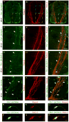
(A–L) Double immunostaining for Flk1 (A,D,G,J: green) and Robo3 (B,E,H,K: red) in E13 rat embryo sections using two different anti-Flk1 antibodies known to label Flk1 in neurons (#SC6251 (AI) and #SC504 (JL)), showing Flk1 expression in pre- and post-crossing commissural axons; panels C,F,I,L show the merged images. Panels DF and GI are higher magnification of the blue and white insets, respectively, shown in AC. The arrows (D,F,G,I,J,L) point to Flk1+ commissural axons. (M–R) Double immunostaining for Flk1 (green in M,P) and Robo3 (red in N) or TAG-1 (red in Q) in dissociated commissural neurons showing expression of Flk1 in the cell body, axon and growth cone of commissural neurons; panels O,R: merged images. Scale bars: AL, 50 μm; MR, 10 μm.
Figure 4
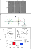
Figure 4. VEGF induces commissural axon turning in a Flk1-dependent manner
(A) Representative images of commissural neurons subjected to a control gradient (BSA, upper panels) or a VEGF gradient (25 ng/ml, lower panels) showing no change of direction in commissural axons exposed to the control gradient (red asterisks in upper panels), but a significant turning towards increasing concentrations of VEGF in neurons exposed to a VEGF gradient (red asterisks in lower panels) over the course of 1.5 hour. Increasing gradient concentrations (from bottom to top) are represented by a wedge. (B) Trajectory plots of a sample of 20 axons in control (left) or 16 axons in 25 ng/ml VEGF (right) gradient. All trajectories have been rotated so that the gradient increases along the y-axis. The initial axon position is shown in black and the axon growth over 1.5 hour is coloured according to the angle turned (scale is shown on the right). (C,D) Scatter plots of the angle turned versus initial angle for commissural axons in a control or a VEGF gradient (25 ng/ml in the outer well). (E) Histogram representing the mean angle turned (±SEM) for initial angles >20° in response to a control gradient (black bar), a VEGF gradient (25 ng/ml; red bar), a VEGF gradient (25 ng/ml) in the presence of anti-Flk1 (100 ng/ml) (white bar), or a VEGF gradient (25 ng/ml) in the presence of anti-Npn1 (10 μg/ml) (blue bar) (one way ANOVA with Bonferronni’s post-test, ***P<0.001; n.s.: not significant). Scale bars: A, 20 μm.
Figure 5
Figure 5. I nactivation of Flk1 causes commissural axon guidance defects in vivo
(A–F) Robo3 immunostaining (red) in embryos with selective inactivation of Flk1 in the dorsal spinal cord (Flk1CN-ko; generated by crossing Wnt-1-Cre with Flk1lox/LacZ mice) and in control embryos (Flk1CN-wt). Compared to Flk1CN-wt embryos (A,D), commissural axons in Flk1CN-ko embryos are defasciculated and some axons project near the lateral edge of the spinal cord and invade the motor columns (white arrows) (B,C,E,F). Panels DF are higher magnification of the insets in AC. Panels B,E and C,F are representative images from two different mutant embryos. (G) Scheme depicting the observed phenotype. Left: normal commissural axons (red) project from the dorsal spinal cord to the floor plate (blue) in Flk1CN-wt embryos. Right: in Flk1CN-ko embryos, axons project in a highly disorganized and defasciculated manner and invade the motor columns. (H) Left: histogram shows the quantification of the area occupied by commissural axons (% of the total spinal cord area). *: P=0.03, Student’s t-test; N=5 Flk1CN-wt; N=8 Flk1CN-ko. Right: histogram showing the penetrance of the phenotype (%); N=13 Flk1CN-wt; N=12 Flk1CN-ko. Differences in the penetrance of the phenotype between Flk1CN-wt and VegfFP-wt might be due to their different genetic background. Scale bars: AC, 10 μm; DF, 20 μm.
Figure 6
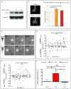
Figure 6. VEGF- induced growth cone turning requires SFKs activation
(A) Immunoblot for anti-phospho-SFK (Y418) (upper panel) and total SFK (lower panel) of dissociated rat commissural neurons incubated with 0, 10 or 25 ng/ml of VEGF for 30 min. (B) Dissociated rat commissural neurons were incubated with vehicle (control) (upper panel) or 10 ng/ml of VEGF (lower panel) for 30 min, and then fixed and immunostained with anti-phospho-SFK (Y418). Dotted lines delineate growth cones. VEGF stimulation leads to an increase in phospho-SFKs in the growth cone. Graph: mean relative levels (± SEM) of phospho-SFK fluorescence in control and VEGF-stimulated (10 or 25 ng/ml) growth cones. The average phospho-SFK fluorescence signal was measured for each growth cone and normalized to the mean signal in control-stimulated growth cones. N=129 for control, N=167 for VEGF (10 ng/ml) and N=107 for VEGF (25 ng/ml), were measured in two independent experiments. *** P<0.0001; * P<0.05 (unpaired t-test). (C) Axons of dissociated commissural neurons in a 25 ng/ml VEGF gradient in a Dunn chamber, in the presence of bath-applied PP2 (0.8 μM) or PP3 (0.8 μM). Inhibition of SFK activity by PP2 inhibits VEGF-mediated turning. PP3 did not inhibit the ability of axons to turn up a VEGF gradient. Black arrow head points to the initial position of the growth cone; white arrowhead points to its final position. (D–F) Scatter plot of the angle turned versus initial angle (D,E) and mean angle turned (F) (± SEM) for initial angles > 20°, show that SFK inhibition by PP2 (E,F), but not by PP3 (D,F), inhibits commissural axon turning towards VEGF (P<0.05; P=0.73, respectively, in unpaired t-test). Axon turning up a VEGF gradient is not perturbed by PP3 and was significantly different from the control or the PP2 condition (P<0.05, initial angle >20°); one way ANOVA with Bonferronni’s multiple comparison post-test, *P<0.05; n.s.: not significant. Scale bars: B, 10 μm, C, 20 μm.

Comment in

References

    1. Avantaggiato V, Orlandini M, Acampora D, Oliviero S, Simeone A. Embryonic expression pattern of the murine figf gene, a growth factor belonging to platelet-derived growth factor/vascular endothelial growth factor family. Mech Dev. 1998;73:221–224. - PubMed
    1. Baldwin ME, Catimel B, Nice EC, Roufail S, Hall NE, Stenvers KL, Karkkainen MJ, Alitalo K, Stacker SA, Achen MG. The specificity of receptor binding by vascular endothelial growth factor-d is different in mouse and man. J Biol Chem. 2001;276:19166–19171. - PubMed
    1. Bellon A, Luchino J, Haigh K, Rougon G, Haigh J, Chauvet S, Mann F. VEGFR2 (KDR/Flk1) signaling mediates axon growth in response to semaphorin 3E in the developing brain. Neuron. 2010;66:205–219. - PubMed
    1. Bocker-Meffert S, Rosenstiel P, Rohl C, Warneke N, Held-Feindt J, Sievers J, Lucius R. Erythropoietin and VEGF promote neural outgrowth from retinal explants in postnatal rats. Invest Ophthalmol Vis Sci. 2002;43:2021–2026. - PubMed
    1. Carmeliet P, Ferreira V, Breier G, Pollefeyt S, Kieckens L, Gertsenstein M, Fahrig M, Vandenhoeck A, Harpal K, Eberhardt C, et al. Abnormal blood vessel development and lethality in embryos lacking a single VEGF allele. Nature. 1996;380:435–439. - PubMed

Publication types

MeSH terms