. 2011 Oct;25(10):1610-9.
doi: 10.1038/leu.2011.120. Epub 2011 Jun 10.

Identification of novel myeloma-specific XBP1 peptides able to generate cytotoxic T lymphocytes: a potential therapeutic application in multiple myeloma

Affiliations

Identification of novel myeloma-specific XBP1 peptides able to generate cytotoxic T lymphocytes: a potential therapeutic application in multiple myeloma

J Bae et al. Leukemia. 2011 Oct.

Abstract

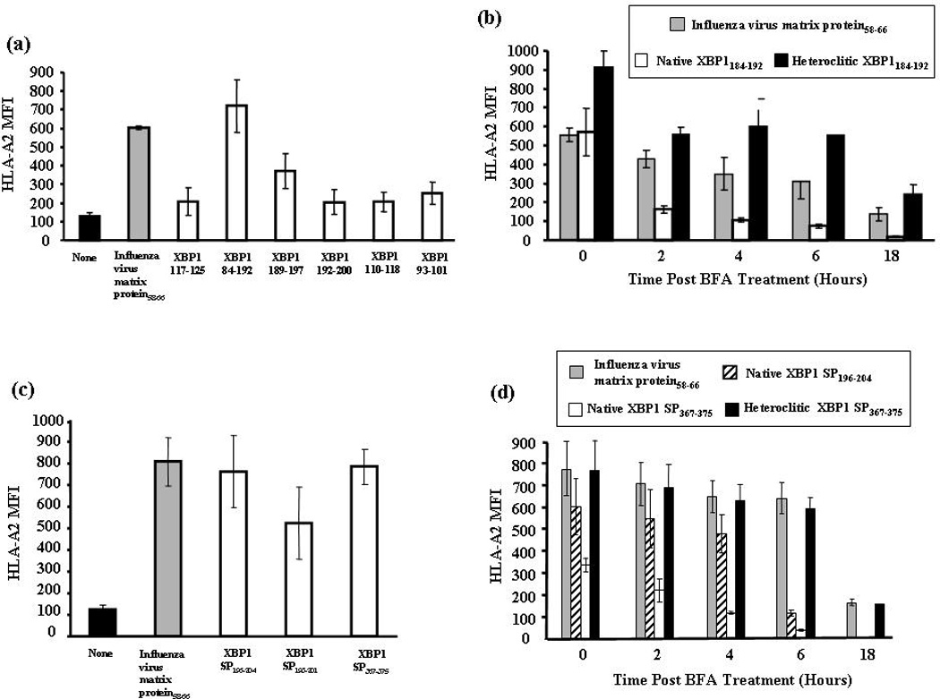
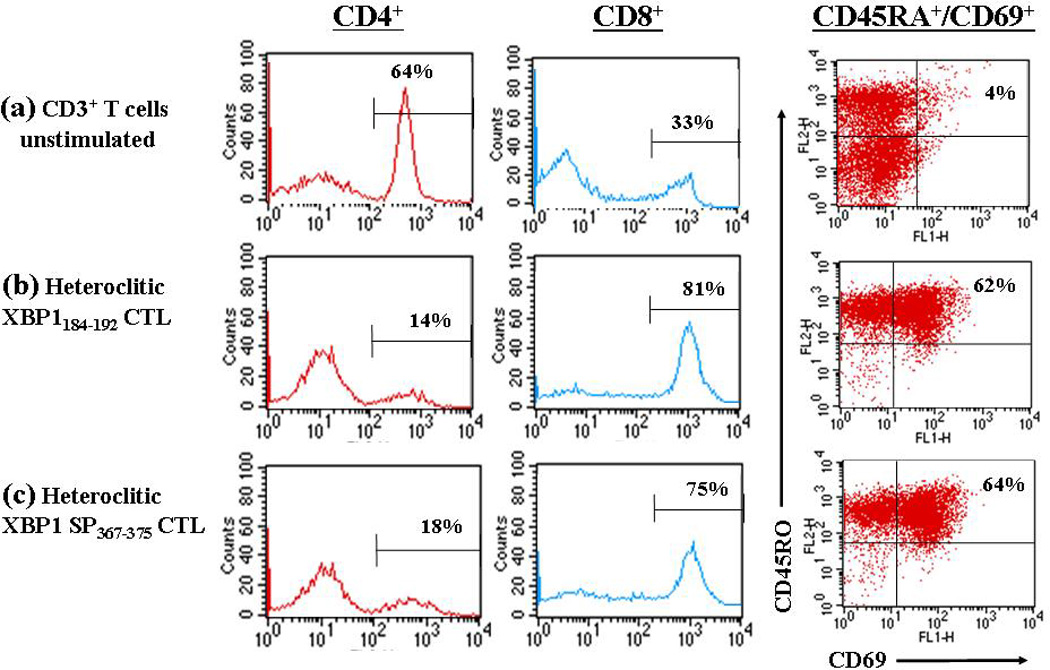
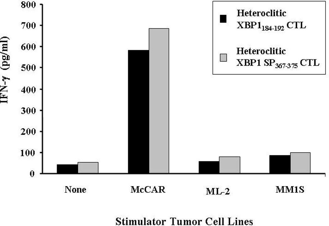
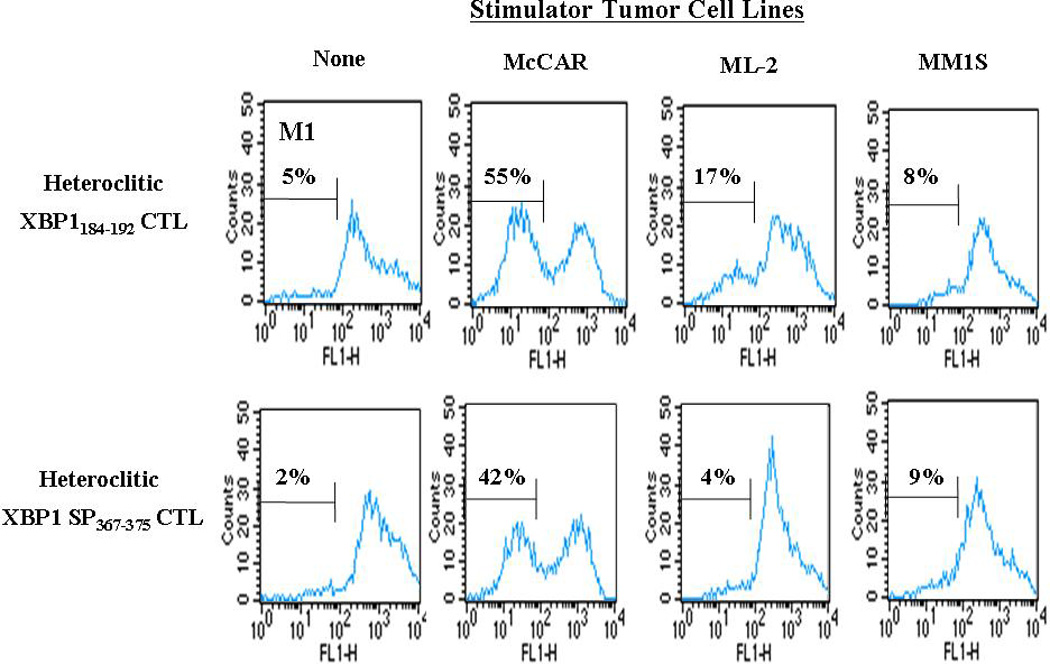
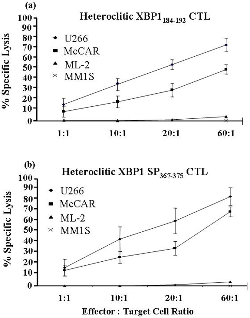
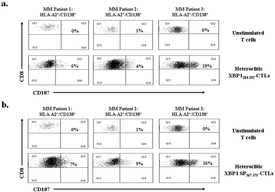
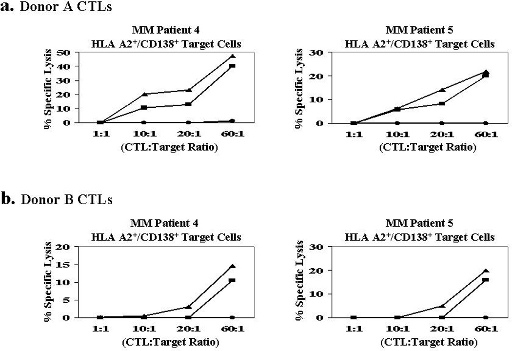
The purpose of these studies was to identify human leukocyte antigen (HLA)-A2(+) immunogenic peptides derived from XBP1 antigens to induce a multiple myeloma (MM)-specific immune response. Six native peptides from non-spliced XBP1 antigen and three native peptides from spliced XBP1 antigen were selected and evaluated for their HLA-A2 specificity. Among them, XBP1(184-192), XBP1 SP(196-204) and XBP1 SP(367-375) peptides showed the highest level of binding affinity, but not stability to HLA-A2 molecules. Novel heteroclitic XBP1 peptides, YISPWILAV or YLFPQLISV, demonstrated a significant improvement in HLA-A2 stability from their native XBP1(184-192) or XBP1 SP(367-375) peptide, respectively. Cytotoxic T lymphocytes generated by repeated stimulation of CD3(+) T cells with each HLA-A2-specific heteroclitic peptide showed an increased percentage of CD8(+) (cytotoxic) and CD69(+)/CD45RO(+) (activated memory) T cells and a lower percentage of CD4(+) (helper) and CD45RA(+)/CCR7(+) (naïve) T cells, which were distinct from the control T cells. Functionally, the cytotoxic T lymphocytes (CTL) demonstrated MM-specific and HLA-A2-restricted proliferation, interferon-γ secretion and cytotoxic activity in response to MM cell lines and importantly, cytotoxicity against primary MM cells. These data demonstrate the distinct immunogenic characteristics of unique heteroclitic XBP1 peptides, which induce MM-specific CTLs and highlights their potential application for immunotherapy to treat the patients with MM or its pre-malignant condition.


Conflict of interest statement

CONFLICT OF INTEREST

There are no competing financial conflicts of interest in relation to the work described.

Figures

Figure 1
Figure 1. Identification of HLA-A2-specific XBP1 peptides and improvement of MHC binding stability by peptide modification
Figure 1a. HLA-A2 binding affinity of non-spliced XBP1 peptides. T2 cells were pulsed overnight with respective XBP1 peptide (50 µg/ml) in serum-fee media. Influenza virus matrix protein58–66 was used as the positive control and T2 cell in media alone as baseline controls in these experiments. Following incubation, T2 cells were harvested, washed, and stained with HLA-A2-FITC mAb for flow cytometric analysis. HLA-A2 binding is shown as an increase in HLA-A2 mean fluorescence intensity (MFI). XBP1184–192 (NISPWILAV) showed the highest HLA-A2 binding affinity among the non-spliced XBP1 peptides. The values represent the mean ± SE of three separate experiments. Figure 1b. HLA-A2 binding stability of non-spliced XBP1 peptides. Native or heteroclitic XBP1 peptide (50 µg/ml)-pulsed T2 cells were washed and incubated with Brefeldin A. At 0, 2, 4, 6, and 18 hours incubation, the cells were stained with HLA-A2-FITC mAb for flow cytometric analysis. Heteroclitic XBP1184–192 (YISPWILAV) peptide showed increased HLA-A2 binding stability compared to the native XBP1184–192 (NISPWILAV) peptide. Binding stability of the heteroclitic peptide was higher than influenza virus matrix protein58–66 (GILGFVFTL), which was used as the HLA-A2-specific positive control peptide. The values represent the mean ± SE of three separate experiments. Figure 1c. HLA-A2 binding affinity of spliced XBP1 peptides. Spliced XBP1 peptides were evaluated for their HLA-A2 binding affinity as described in Figure 1a. XBP1 SP196–204 (GILDNLDPV) and XBP1 SP367–375 (ELFPQLISV) showed the highest HLA-A2 binding affinity among the spliced XBP1 peptides. The values represent the mean ± SE of three separate experiments. Figure 1d. HLA-A2 binding stability of spliced XBP1 peptides. Spliced XBP1 peptides were analyzed for their HLA-A2 binding stability as discussed in Figure 1b. Heteroclitic XBP1 SP367–375 (YLFPQLISV) peptide displayed increased HLA-A2 binding stability compared to its native XBP1 SP367–375 (ELFPQLISV) or another native XBP1 SP196–204 (GILDNLDPV) peptide. The values represent the mean ± SE of three separate experiments.
Figure 2
Figure 2. Distinct phenotype of CTLs stimulated with heteroclitic XBP1-specific peptides
Enriched CD3+ T cells obtained from a normal HLA-A2+ donor were stimulated weekly with irradiated antigen-presenting cells pulsed with respective heteroclitic XBP1 peptide. One week after the 4th cycle of peptide stimulation, the CD3+ T cells were analyzed for effector cells by flow cytometry. The percentage of CD4+ helper T cells was decreased in the T cells stimulated with either (b) heteroclitic XBP1184–192 YISPWILAV) peptide or (c) heteroclitic XBP1 SP367–375 (YLFPQLISV) peptide, compared to (a) no peptide. In contrast, the percentage of CD8+ CTLs and CD69+/CD45RO+ (activated memory) effector cells was increased in the CD3+ T cells stimulated with respective XBP1 peptide, compared to no peptide control. The results are representative of three independent experiments.
Figure 3
Figure 3. HLA-A2 restricted and XBP1-specific IFN-γ secretion by XBP1-CTLs stimulated with heteroclitic peptides
CTLs generated by repeated stimulation (4×) with heteroclitic XBP1184–192 (YISPWILAV) or heteroclitic XBP1 SP367–375 (YLFPQLISV) peptide were incubated with McCAR (MM; HLA-A2+/XBP1+), ML-2 (AML; HLA-A2+/XBP1) or MM1S (MM; HLA-A2/XBP1+) tumor cell lines. After 24 hours of incubation, the supernates were collected and IFN-γ secretion was measured by ELISA. An increase in IFN-γ secretion was detected in both XBP1-CTLs cultures in response to McCAR (HLA-A2+/XBP1+), but not to ML-2 (HLA-A2+/XBP1) or MM1S (HLA-A2/XBP1+) cells. XBP1-CTLs alone were used to examine the background IFN-γ release. The results are representative of three independent experiments.
Figure 4
Figure 4. Induction of HLA-A2-restricted and XBP1-specific cell proliferation of XBP1-CTLs in response to multiple myeloma cells
Proliferation of CFSE-labeled XBP1-CTLs was measured in response to antigen stimulation by flow cytometry on day 4 of culture. The cell proliferation of heteroclitic XBP1184–192 (YISPWILAV) and heteroclitic XBP1 SP367–375 (YLFPQLISV) CTLs were both XBP1 antigen-specific and HLA-A2-restricted in response to the McCAR (HLA-A2+/XBP1+) myeloma cell line. The CTLs did not proliferate in response to either the XBP1 antigen or MHC mismatched cell lines ML-2 (HLA-A2+/XBP1) or MM1S (HLA-A2/XBP1+), respectively. Background proliferation was determined using CFSE labeled heteroclitic XBP1-CTLs cultured in media alone. The results are representative of three independent experiments.
Figure 5
Figure 5. HLA-A2-restricted and antigen-specific cytotoxicity of the XBP1-CTLs
The specific cytotoxic activities of the heteroclitic XBP1-CTLs were analyzed one week after their 4th stimulation using the calcein cytotoxicity assay. A high level of cytotoxic activity against U266 (MM, HLA-A2+/XBP1+) (♦) and McCAR (MM, HLA-A2+/XBP1+) (■), but not against the ML-2 (AML, HLA-A2+/XBP1) (▲) and MM1S (HLA-A2/XBP1+) (X) cell lines was demonstrated by both the (a) heteroclitic XBP1184–192 CTLs and (b) heteroclitic XBP1 SP367–375 -CTLs. The values represent the mean±SE of three separate experiments.
Figure 6
Figure 6. XBP1-CTL degranulation in response to primary HLA-A2+/CD138+ patient MM cells
Degranulation of heteroclitic XBP1-CTLs was analyzed by flow cytometry one week after their 4th stimulation, using CD107a, a surface antigen transiently present on the cell surface after release of cytotoxic granules. CD107a expression on (a) heteroclitic XBP1184–192-CTLs and (b) heteroclitic SP367–375–CTLs following stimulation with primary CD138+ MM cells from three HLA-A2+ patients. Control unstimulated T cells did not degranulate when exposed to the primary HLA-A2+/ CD138+ MM cells. The results are expressed as the percent CD107a of CD8+ T cells.
Figure 7
Figure 7. Cytotoxicity of XBP1-CTLs against primary HLA-A2+/CD138+ patient MM cells
The specific cytotoxic activity of heteroclitic XBP1-CTLs were analyzed one week after their 4th stimulation using the calcein-release cytotoxicity assay. Heteroclitic XBP1184–192 CTLs (■) and heteroclitic SP367–375–CTLs (▲) generated from T cells of (a) Donor 1 and (b) Donor 2 demonstrate a high level of cytotoxic activity against primary CD138+ MM cells from two HLA-A2+ MM patients. Control unstimulated T cells (●) did not lyse primary CD138+ MM cells from the HLA-A2+ MM patients.

References

    1. Kumar S. Multiple myeloma - current issues and controversies. Cancer Treat Rev. 2010;36(Suppl 2):S3–S11. - PubMed
    1. Laubach JP, Richardson PG, Anderson KC. The evolution and impact of therapy in multiple myeloma. Med Oncol. 2010;27(Suppl 1):S1–S6. - PubMed
    1. Jagannath S, Kyle RA, Palumbo A, Siegel DS, Cunningham S, Berenson J. The current status and future of multiple myeloma in the clinic. Clin Lymphoma Myeloma Leuk. 2010;10:28–43. - PubMed
    1. Bensinger WI. Role of autologous and allogeneic stem cell transplantation in myeloma. Leukemia. 2009;23:442–448. - PMC - PubMed
    1. Zeiser R, Finke J. Allogeneic haematopoietic cell transplantation for multiple myeloma: reducing transplant-related mortality while harnessing the graft-versusmyeloma effect. Eur J Cancer. 2006;42:1601–1611. - PubMed

Publication types

MeSH terms