

In vivo confocal microscopy of the corneal endothelium: comparison of three morphometry methods after corneal transplantation
- PMID: 21660067
- PMCID: PMC3178261
- DOI: 10.1038/eye.2011.121
In vivo confocal microscopy of the corneal endothelium: comparison of three morphometry methods after corneal transplantation
Abstract
Purpose: The purpose of this study was to assess the endothelium of corneal grafts by in vivo confocal microscopy (IVCM), and to evaluate an automated endothelial software system in comparison with a manual cell count and planimetry.
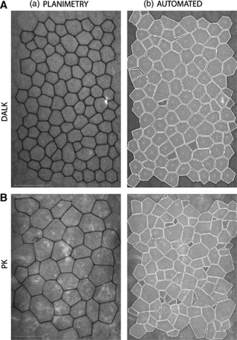
Patients and methods: Overall, 40 corneal grafts (20 deep anterior lamellar keratoplasties (DALKs) and 20 penetrating keratoplasties (PKs)) were assessed by scanning-slit IVCM. The endothelial cell density (ECD) was estimated with the automated and the manual cell count method of the instrument's Nidek Advanced Vision Information System (NAVIS) software. The results were compared with planimetry as the reference method, and the agreement was assessed.
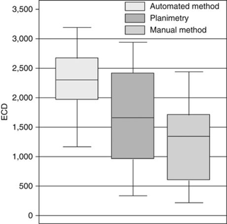
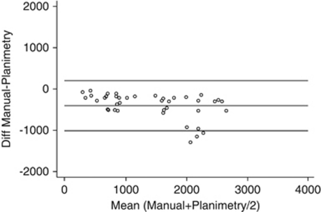
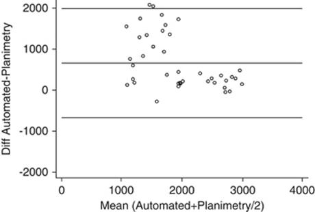
Results: The mean (±SD) automated ECD was 2278±524 cells/mm(2) (range 1167-3192 cells/mm(2)), whereas the manual cell count method gave significantly lower ECDs with a mean of 1213±677 cells/mm(2) (range 218-2440 cells/mm(2); P<0.001). The manual cell counts were also significantly lower than those by planimetry, with a mean ECD of 1617±813 cells/mm(2) (range 336-2941, P<0.001). Bland-Altman analyses indicated that the limits of agreement (LoA) between the automated and the planimetry method were -671 and +1992 cells/mm(2), whereas they were -1000 and +202 cells/mm(2) when comparing the manual cell counts with planimetry.
Conclusion: Following keratoplasty, the NAVIS automated method is likely to overestimate endothelial cell counts due to oversegmenting of the cell domains. Automated ECDs are substantially higher than those by the manual counting method or planimetry. The differences are considerably larger post-keratoplasty than for normal corneas, and the methods should not be used interchangeably.
Figures
References
-
- Reinhart WJ, Musch DC, Jacobs DS, Lee WB, Kaufman SC, Shtein RM. Deep anterior lamellar keratoplasty as an alternative to penetrating keratoplasty: a report by the American Academy of Ophthalmology. Ophthalmology. 2011;118 (1:209–218. - PubMed
-
- Bourne WM. Cellular changes in transplanted human corneas. Cornea. 2001;20 (6:560–569. - PubMed
-
- Inoue K, Kimura C, Amano S, Oshika T, Tsuru T. Corneal endothelial cell changes twenty years after penetrating keratoplasty. Jpn J Ophthalmol. 2002;46 (2:189–192. - PubMed
-
- Patel SV, Hodge DO, Bourne WM. Corneal endothelium and postoperative outcomes 15 years after penetrating keratoplasty. Am J Ophthalmol. 2005;139 (2:311–319. - PubMed
Publication types
MeSH terms
LinkOut - more resources
Full Text Sources
Miscellaneous
