

Be careful... She has a pituitary gland in her nose
- PMID: 21660520
- PMCID: PMC3505508
- DOI: 10.1007/s11102-011-0320-5
Be careful... She has a pituitary gland in her nose
Abstract
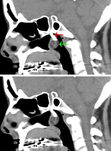
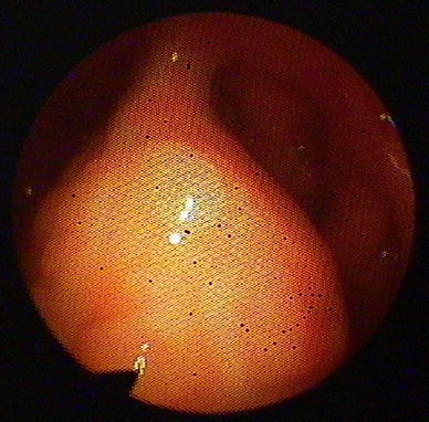
In this case report we describe a 38 year-old-female with galactorrhea several months after the birth of an anencephalic child. She had hyperpolactemia and imaging of the pituitary gland revealed a midline defect and a nasopharyngeal mass compatible with a meningo-(hypophyso-) encephalocele and possibly an ectopic teratoma or desmoid. She was treated with dopamine agonists for 10 years and after cessation of therapy her prolactin levels remain normal. The nasopharyngeal mass remained unchanged over time and there were no signs of hypopituitarism. The hyperprolactinemia at presentation was probably caused by earlier pregnancy and stalk dysfunction due to traction by the mass. With decline of pituitary size, after starting dopamine agonists, the traction probably reduced resulting in a normal prolactin level. Our patient was warned against manipulation in de nose, because this could damage the meningo-encephalocele. An MRI will be preformed every 2 years to monitor changes in de mass.
Figures

Similar articles
-
Macroprolactinemia in a patient with infertility and hyperprolactinemia.South Med J. 2006 Nov;99(11):1282-4. doi: 10.1097/01.smj.0000232973.76521.5d. South Med J. 2006. PMID: 17195425
-
Del Castello syndrome--an unusual presentation.J Indian Med Assoc. 2002 Aug;100(8):524, 526. J Indian Med Assoc. 2002. PMID: 12675188
-
Clinical presentation of hyperprolactinemia.J Reprod Med. 1999 Dec;44(12 Suppl):1085-90. J Reprod Med. 1999. PMID: 10649815
-
Hyperprolactinemia: etiology, diagnosis, and management.Semin Reprod Med. 2002 Nov;20(4):365-74. doi: 10.1055/s-2002-36709. Semin Reprod Med. 2002. PMID: 12536359 Review.
-
[Current diagnosis and treatment of hyperprolactinemia].Rev Med Inst Mex Seguro Soc. 2016 Jan-Feb;54(1):111-21. Rev Med Inst Mex Seguro Soc. 2016. PMID: 26820213 Review. Spanish.
References
-
- Yoshimoto Y, Noguchi M, Tsutsumi Y. A case of transethmoidal encephalocele. No Shinkei Geka. 1992;20:249–254. - PubMed
Publication types
MeSH terms
Substances
LinkOut - more resources
Full Text Sources
