Diffusion-weighted MRI reveals epiphyseal and metaphyseal abnormalities in Legg-Calvé-Perthes disease: a pilot study
- PMID: 21660596
- PMCID: PMC3171554
- DOI: 10.1007/s11999-011-1931-x
Diffusion-weighted MRI reveals epiphyseal and metaphyseal abnormalities in Legg-Calvé-Perthes disease: a pilot study
Abstract
Background: Legg-Calvé-Perthes disease (LCP) is thought to be associated with ischemic events in the femoral head. However, the types and patterns of reperfusion after these ischemic events are unclear.
Purposes: We therefore determined whether (1) there would be any age-related diffusion changes; (2) diffusion-weighted MR imaging would reveal ischemic damage; and (3) diffusion changes are correlated with prognostic MR findings in patients with LCP.
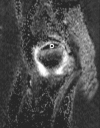
Methods: We prospectively performed conventional, perfusion, and diffusion-weighted MR imaging studies in 17 children with unilateral LCP. We then measured the apparent diffusion coefficient (ADC) values in the epiphysis and the metaphysis, and compared them with those of the contralateral normal side. Based on perfusion MR imaging, we assessed reperfusion to the epiphysis as either periphyseal or transphyseal. We studied T2-signal intensity changes in the metaphysis and the presence of focal physeal irregularity. We correlated diffusion changes with reperfusion to the epiphysis, T2-signal intensity change, and focal physeal irregularity.
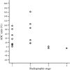
Results: Normal diffusion decreased with age. In LCP hips, epiphyseal diffusion increased early and remained elevated through the healing stage. Six of the 17 patients who had a metaphyseal ADC greater than 50% over the normal side had 13 times greater odds of having an association with transphyseal reperfusion to the epiphysis. The increase of metaphyseal ADC also was associated with an increased T2-signal intensity in the metaphysis and presence of focal physeal irregularity.
Conclusions: Diffusion-weighted MR imaging can be used as a complimentary modality to evaluate ischemic tissue damage with a potential prognostic value in patients with LCP.
Figures


References
-
- Baur A, Stabler A, Bruning R, Bartl R, Krodel A, Reiser M, Deimling M. Diffusion-weighted MR imaging of bone marrow: differentiation of benign versus pathologic compression fractures. Radiology. 1998;207:349–356. - PubMed
-
- Catterall A. The natural history of Perthes’ disease. J Bone Joint Surg Br. 1971;53:37–53. - PubMed
MeSH terms
LinkOut - more resources
Full Text Sources

