. 2011 Sep;226(9):2360-9.
doi: 10.1002/jcp.22568.

RASSF1A suppresses melanoma development by modulating apoptosis and cell-cycle progression

Affiliations

RASSF1A suppresses melanoma development by modulating apoptosis and cell-cycle progression

Mei Yi et al. J Cell Physiol. 2011 Sep.

Abstract

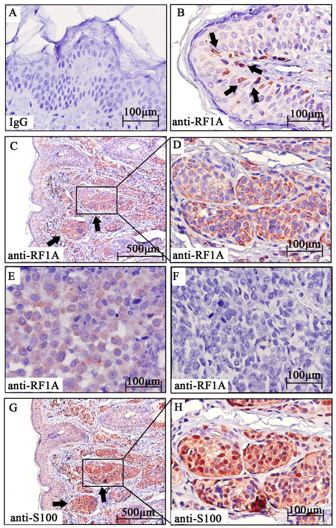
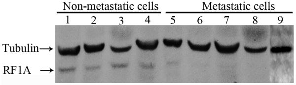
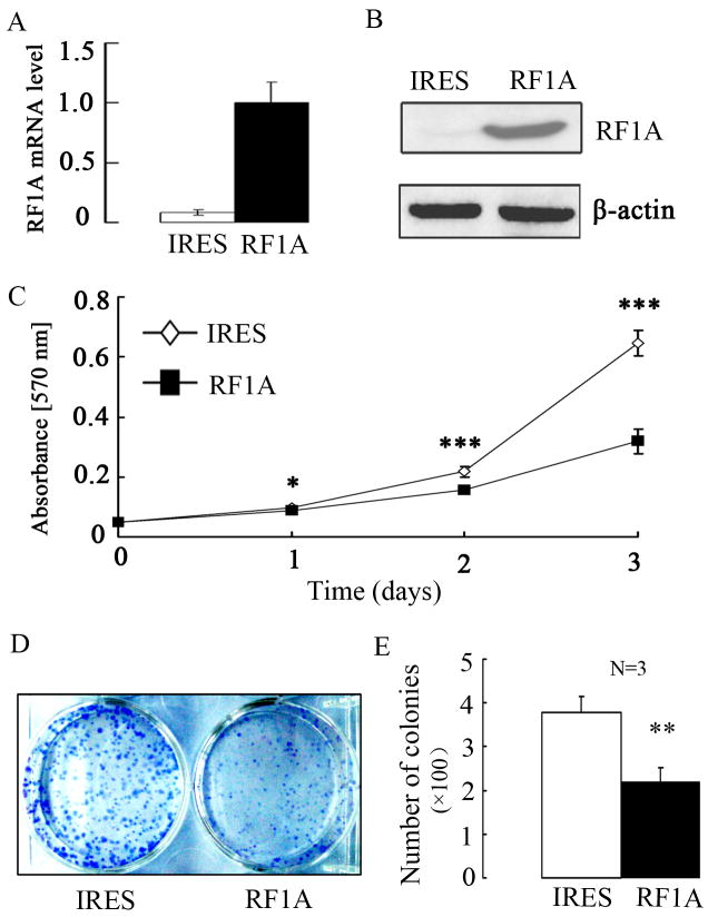
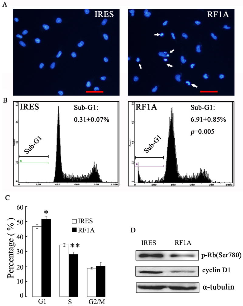
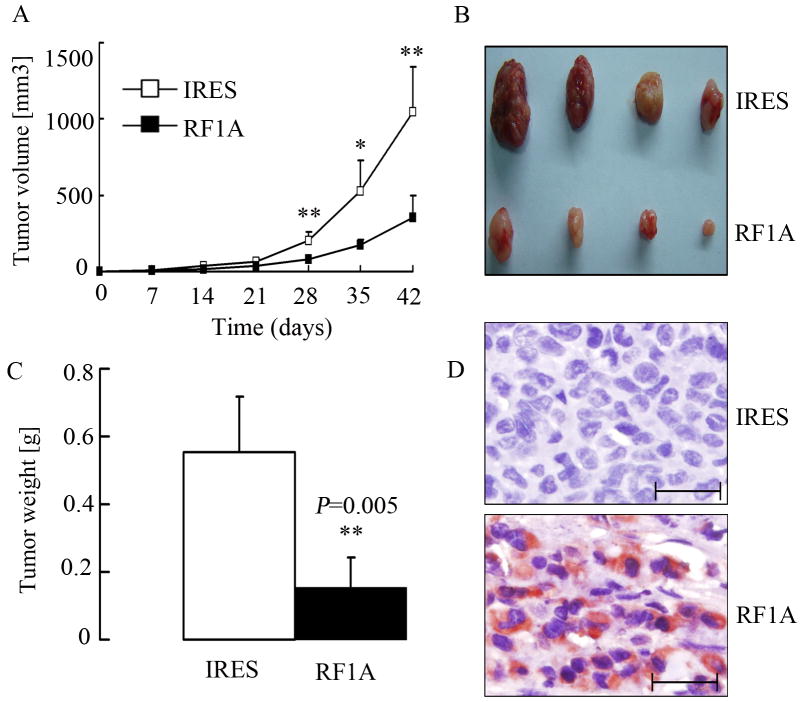
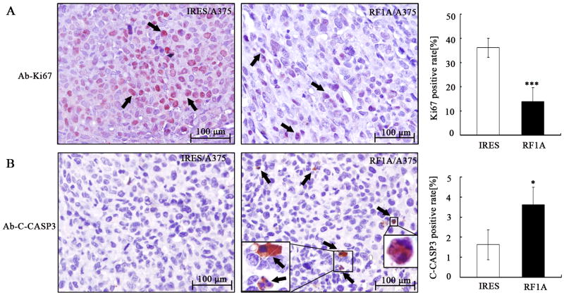
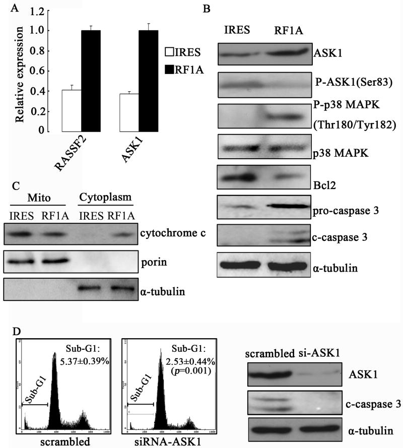
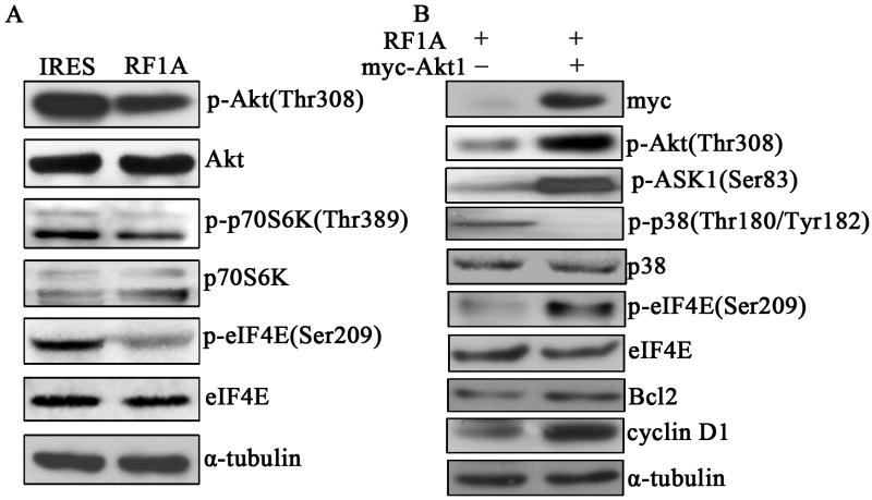
The tumor suppressor candidate gene Ras association domain family 1, isoform A (RASSF1A) encodes a microtubule-associated protein that is implicated in the regulation of cell proliferation, migration, and apoptosis. Several studies indicate that down-regulation of RASSF1A resulting from promoter hypermethylation is a frequent epigenetic abnormality in malignant melanoma. In this study, we report that compared with melanocytes in normal skins or benign skin lesions, RASSF1A is down-regulated in melanoma tissues as well as cell lines, and its expression negatively correlates with lymph node metastasis. Following ectopic expression in RASSF1A-deficient melanoma A375 cell line, RASSF1A reduces cell viability, suppresses cell-cycle progression but enhances apoptotic cell death. In vivo, RASSF1A expression inhibits the tumorigenic potential of A375 cells in nude mice, which also correlates with decreased cell proliferation and increased apoptosis. On the molecular level, ectopic RASSF1A expression leads to differential expression of 209 genes, including 26 down-regulated and 183 up-regulated ones. Among different signaling pathways, activation of the apoptosis signal-regulating kinase 1 (ASK1)/p38 MAP kinase signaling is essential for RASSF1A-induced mitochondrial apoptosis, and the inhibition of the Akt/p70S6 kinase/eIF4E signaling is also important for RASSF1A-mediated apoptosis and cell-cycle arrest. This is the first study exploring the biological functions and the underlying mechanisms of RASSF1A during melanoma development. It also identifies potential targets for further diagnosis and clinical therapy.


Conflict of interest statement

Competing interests: The authors declare no competing interests.

Figures

Figure 1
Figure 1. RASSF1A is down-regulated in MM samples
(A). Staining of normal skin with normal mouse IgG as the primary antibody, which served as negative control for immunohistochemical staining. (B). Strong positive cytoplasmic staining of RASSF1A protein (red) in melanocytes (arrowhead) within normal skin. (C). Low-power magnification of nevus pigmentosus tissue stained with RASSF1A, which showed strong positive cytoplasmic staining of RASSF1A protein in nevus nest (arrowhead). (D). High-power magnification of the squared field from (C). (E). Weak positive staining of RASSF1A protein in melanoma tissues without lymph node metastasis. (F). Absence of RASSF1A protein in primary melanoma tissues with lymph node metastasis. (G). Low-power magnification of S100 staining in nevus pigmentosus, which showed strong positive nuclear and cytoplasmic signal (red) in nevus nest (arrowhead). (H). High-power magnification of the squared field from (G).
Figure 2
Figure 2. RASSF1A is down-regulated in MM cell lines
Western blot analysis of RASSF1A in various melanoma cell lines. α-Tubulin was used as a loading control. 1: WM1552C; 2:WM1341D; 3: WM793; 4: WM164; 5:1205Lu; 6: MeWo; 7: A375SM; 8: M14; 9: A375.
Figure 3
Figure 3. Exogenous expression of RASSF1A suppresses in vitro cell viability
(A). The expression of RASSF1A mRNA was analyzed by qRT-PCR in stable RASSF1A (RF1A) and control (IRES) cell lines. GAPDH was used as internal control. (B). The exogenous expression of RASSF1A protein was analyzed by Western blot analysis. β-Actin was used as a loading control. (C). For growth curves, RASSF1A and control cells were seeded into 96-well plates, with cell viability at indicated time points determined by MTT assay. Data are presented as the mean±SEM of values from 3 independent experiments. (D). A typical microphotograph of colony derived from RASSF1A and control cells. (E). Quantification of the colonies formed in colony formation assay. Data are presented as the mean±SEM of values from 3 independent experiments. * P<0.05, ** P<0.01, and *** P<0.001, as compared with control cells.
Figure 4
Figure 4. Exogenous expression of RASSF1A induces apoptotic cell death and G1-S cell cycle arrest in A375 cells
(A). Respresentative microphotographs of Hoechst33258 staining showed exogenous expression of RASSF1A enhanced apoptotic cell death in A375 cells. IRES, control cells; RF1A, RASSF1A cells. The arrows indicated apoptotic cells, which showed condensed DNA. Scale bar, 50 μm. (B). Representative cell-cycle distribution analysis by FACS on RASSF1 and control cells. The percentage of sub-G1 population was indicated. (C). Quantification of three independent cell-cycle distribution analysis as performed in (B) * P < 0.05, as compared to control cells. (D). The expressions of cyclin D1 and phospho-Rb were analyzed by Western blot analysis. α-tubulin was used as an internal control.
Figure 5
Figure 5. Exogenous expression of RASSF1A suppresses in vivo tumorigenesis
(A). Tumor growth curve. Control (IRES) and RASSF1A (RF1A) cells were injected s.c. into BALB/c nude mice (N=4/group), and tumors were monitored every 7 days with the volume calculated. * P<0.05, ** P<0.01, as compared to tumors from control cells. (B). Photograph of tumors after isolation. (C). The wet weight of tumors was measured after excision. * P<0.01, as compared to tumors from control cells. (D). Tumors were subjected to immunohistochemistry assays for the expression of RASSF1A protein, which showed strong positive staining (red) in tumors from RASSF1A cells. Scale bar, 50 μm.
Fig. 6
Fig. 6. Exogenous expression of RASSF1A suppresses cell proliferation and induces apoptosis in vivo
Xenograft tumors derived from control (IRES) and RASSF1A (RF1A) cells (N=4/group) were subjected to immunohistochemical analysis on Ki67 (A) and cleaved casp3 (B) for assessing proliferation and apoptosis, respectively. . Arrows indicate positively stained cells. *** P < 0.001; * P < 0.05, as compared to tumors from control cells.
Figure 7
Figure 7. Exogenous expression of RASSF1A mediates mitochondrial apoptosis through up-regulation of ASK1
(A). qRT-PCR of RASSF2 and ASK1 expression in control (IRES) or RASSF1A (RF1A) cells. GAPDH was used as internal control. (B). The expressions of ASK1, phospho-ASK1, p38, phospho-p38, Bcl2, caspase 3 and cleaved-caspase 3 were analyzed by Western blotting. (C). The subcellular distribution of cytochrome c was analyzed by Western blot. (D). Apoptosis in RASSF1A cells transfected with either control (scrambled) or ASK1-specific siRNA (siRNA-ASK1) was determined by FACS (left two panels, sub-G1 population) or Western blotting on cleaved casp-3 (right panel). Representative images from three independent experiments are shown.
Figure 8
Figure 8. Exogenous expression of RASSF1A inhibits Akt/p70S6K/eIF4E signaling pathway
(A). The expressions of phospho-Akt (p-Akt), Akt, p-p70S6K, p70S6K, p-eIF4E, and eIF4E were analyzed by Western blottingin control (IRES) and RASSF1A (RF1A) cells. (B). The expressions of myc, p-Akt, p-ASK1, p-p38, p38, p-eIF4E, eIF4E, Bcl2 and cyclin D1 were determined by Western blotting in RASSF1A cells transfected with either empty (-) or Akt1-expressing vector (+). α-tubulin was used as an internal control.A single representative from three independent experiments is shown.

References

    1. Burbee DG, Forgacs E, Zochbauer-Muller S, Shivakumar L, Fong K, Gao B, Randle D, Kondo M, Virmani A, Bader S. Epigenetic inactivation of RASSF1A in lung and breast cancers and malignant phenotype suppression. JNCI Journal of the National Cancer Institute. 2001;93(9):691. - PMC - PubMed
    1. Chow LSN, Lo KW, Kwong J, To KF, Tsang KS, Lam CW, Dammann R, Huang DP. RASSF1A is a target tumor suppressor from 3p21. 3 in nasopharyngeal carcinoma. International Journal of Cancer. 2004;109(6):839–847. - PubMed
    1. Dammann R, Li C, Yoon JH, Chin PL, Bates S, Pfeifer GP. Epigenetic inactivation of a RAS association domain family protein from the lung tumour suppressor locus 3p21. 3. Nature genetics. 2000;25(3):315–319. - PubMed
    1. Goto Y, Ferrone S, Arigami T, Kitago M, Tanemura A, Sunami E, Nguyen SL, Turner RR, Morton DL, Hoon DSB. Human high molecular weight-melanoma-associated antigen: utility for detection of metastatic melanoma in sentinel lymph nodes. Clinical Cancer Research. 2008;14(11):3401. - PMC - PubMed
    1. Hatai T, Matsuzawa A, Inoshita S, Mochida Y, Kuroda T, Sakamaki K, Kuida K, Yonehara S, Ichijo H, Takeda K. Execution of apoptosis signal-regulating kinase 1 (ASK1)-induced apoptosis by the mitochondria-dependent caspase activation. Journal of Biological Chemistry. 2000;275(34):26576. - PubMed

Publication types

MeSH terms

Substances