Comparative Study
. 2011 Aug;44(2):221-34.
doi: 10.1002/mus.22033. Epub 2011 Jun 9.

Acellular nerve allografts in peripheral nerve regeneration: a comparative study

Affiliations
Comparative Study

Acellular nerve allografts in peripheral nerve regeneration: a comparative study

Amy M Moore et al. Muscle Nerve. 2011 Aug.

Abstract

Introduction: Processed nerve allografts offer a promising alternative to nerve autografts in the surgical management of peripheral nerve injuries where short deficits exist.

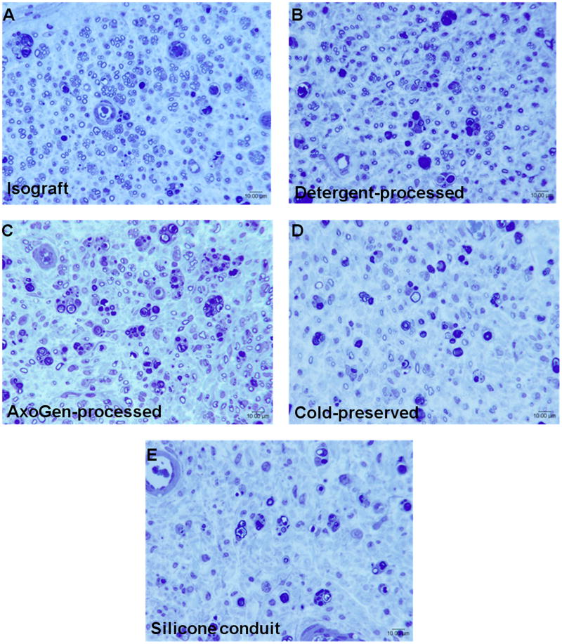
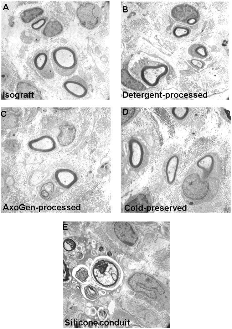
Methods: Three established models of acellular nerve allograft (cold-preserved, detergent-processed, and AxoGen-processed nerve allografts) were compared with nerve isografts and silicone nerve guidance conduits in a 14-mm rat sciatic nerve defect.

Results: All acellular nerve grafts were superior to silicone nerve conduits in support of nerve regeneration. Detergent-processed allografts were similar to isografts at 6 weeks postoperatively, whereas AxoGen-processed and cold-preserved allografts supported significantly fewer regenerating nerve fibers. Measurement of muscle force confirmed that detergent-processed allografts promoted isograft-equivalent levels of motor recovery 16 weeks postoperatively. All acellular allografts promoted greater amounts of motor recovery compared with silicone conduits.

Conclusion: These findings provide evidence that differential processing for removal of cellular constituents in preparing acellular nerve allografts affects recovery in vivo.


Figures

Figure 1
Figure 1
Histomorphometric findings reveal varying degrees of axonal regeneration through fresh nerve isografts, processed nerve allografts, and nerve guidance conduits 6 weeks post-operatively. (A) Fiber counts demonstrate significantly greater numbers of nerve fibers distal to implanted isografts and detergent-processed allografts than AxoGen®-processed allografts and cold-preserved allografts. (B, C) Calculation of percent nerve tissue and nerve fiber density show increased amount of neural tissue distal to implanted isografts and detergent-processed allografts than AxoGen®-processed allografts and cold-preserved allografts, though no statistical differences were observed. (D) Fiber width measurements demonstrate similar degrees of fiber maturation amongst all implanted nerve allografts. Silicone nerve guidance conduits 14 mm in length did not support successful axonal regeneration, precluding histomorphometric analysis. (E) No significant differences were observed in the amount of debris present in the nerve distal to the implanted grafts/conduits. Data represents the mean ± standard deviation; * indicates statistical significance (p < 0.05); # indicates p < 0.05 vs Isograft, Detergent-processed, AxoGen®-processed and Cold-preserved; ˆ indicates p < 0.05 vs Detergent-processed, AxoGen®-processed, Cold-preserved and Conduit.
Figure 2
Figure 2
Representative histological sections demonstrate populations of axons successfully regenerating through fresh nerve isografts, processed nerve allografts, and nerve guidance conduits 6 weeks post-operatively. Sections acquired 3-5 mm distal to implanted nerve isografts (A) and detergent-processed allografts (B) show numerous myelinated axons loosely organized into regenerating units. Sections acquired distal to AxoGen®-processed nerve allografts (C) and cold-preserved nerve allografts (D) show few myelinated axons successfully innervating the host nerve distal to the repair site. Host nerve tissue distal to implanted nerve guidance conduits (E) demonstrate no healthy, myelinated axons.
Figure 3
Figure 3
Representative electron micrographs reveal unmyelinated and remyelinated axons regenerating through fresh nerve isografts, processed nerve allografts, and nerve guidance conduits 6 weeks post-operatively. Micrographs acquired distal to implanted nerve isografts (A), detergent-processed allografts (4360× magnification) (B), AxoGen®-processed nerve allografts (4360× magnification) (C), and cold-preserved nerve allografts (6400× magnification) (D) show normal remyelination of regenerating axons and multiple unmyelinated axons within the host nerve (4360× magnification). Micrographs acquired distal to implanted nerve guidance conduits (E) demonstrate degenerating axons, neural debris, and an absence of healthy, regenerating axons (4360× magnification).
Figure 4
Figure 4
Evoked muscle force measurements reveal differential motor recovery in distal musculature 16 weeks after implantation of fresh nerve isografts, processed nerve allografts, and nerve guidance conduits. (A) Measurements of maximum isometric force production in EDL muscle innervated by repaired sciatic nerve demonstrate the AxoGen®-processed nerve allografts and cold-preserved nerve allografts support significantly lower degrees of motor recovery compared to fresh nerve isografts. In comparison, silicone nerve guidance conduits did not support any functional motor recovery in distal musculature. (B) Assessment of EDL muscle mass shows that muscles innervated by sciatic nerves repaired with either processed nerve allografts experienced similar degrees of atrophy. EDL muscles innervated by sciatic nerves repaired with AxoGen®-processed nerve allografts did exhibit greater degrees of atrophy, though results were not statistically significant. (C) Calculation of maximum specific force production in reinnervated EDL muscle reveals that, upon correction for differences in muscle atrophy, AxoGen®-processed nerve allografts and cold-preserved nerve allografts still support significantly lower degrees of motor recovery compared to fresh nerve isografts. Data represents the mean ± standard deviation; * indicates statistical significance (p < 0.05) compared to nerve isograft.
Figure 5
Figure 5
Representative force recordings obtained from EDL muscle innervated by repaired sciatic nerve show differential recovery of motor function 16 weeks after implantation of fresh nerve isografts, processed nerve allografts, and nerve guidance conduits. Comparison of evoked twitch (A) and tetanic (B) responses demonstrate improved force production in muscles innervated by nerve repaired with nerve isografts and detergent-processed nerve allograft. Observation of normal tetanic responses in EDL muscle innervated by sciatic nerve repaired with both nerve isografts and processed allografts confirm normal function of regenerated motor axons and corresponding motor units.

References

    1. Noble J, Munro CA, Prasad VS, Midha R. Analysis of upper and lower extremity peripheral nerve injuries in a population of patients with multiple injuries. J Trauma. 1998;45(1):116–122. - PubMed
    1. Bain JR, Mackinnon SE, Hudson AR, Falk RE, Falk JA, Hunter DA. The peripheral nerve allograft: an assessment of regeneration across nerve allografts in rats immunosuppressed with cyclosporin A. Plastic and reconstructive surgery. 1988;82(6):1052–1066. - PubMed
    1. Bain JR, Mackinnon SE, Hudson AR, Falk RE, Falk JA, Hunter DA. The peripheral nerve allograft: a dose-response curve in the rat immunosuppressed with cyclosporin A. Plastic and reconstructive surgery. 1988;82(3):447–457. - PubMed
    1. Strasberg SR, Hertl MC, Mackinnon SE, Lee CK, Watanabe O, Tarasidis G, Hunter DA, Wong PY. Peripheral nerve allograft preservation improves regeneration and decreases systemic cyclosporin A requirements. Exp Neurol. 1996;139(2):306–316. - PubMed
    1. Midha R, Mackinnon SE, Evans PJ, Best TJ, Hare GM, Hunter DA, Falk-Wade JA. Comparison of regeneration across nerve allografts with temporary or continuous cyclosporin A immunosuppression. J Neurosurg. 1993;78(1):90–100. - PubMed

Publication types