Review
. 2011:65:261-86.
doi: 10.1146/annurev-micro-121809-151631.

Ligand-binding PAS domains in a genomic, cellular, and structural context

Affiliations
Review

Ligand-binding PAS domains in a genomic, cellular, and structural context

Jonathan T Henry et al. Annu Rev Microbiol. 2011.

Abstract

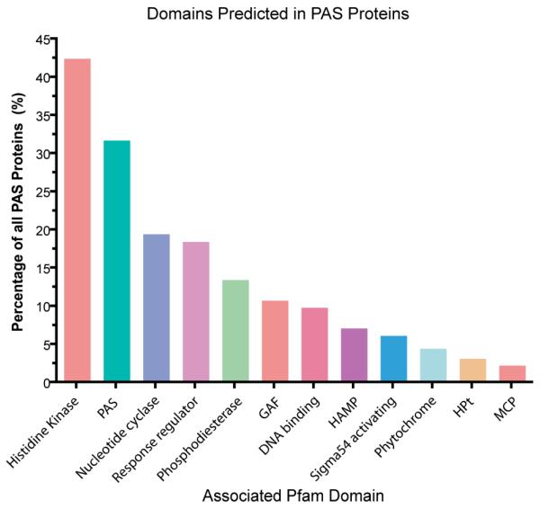
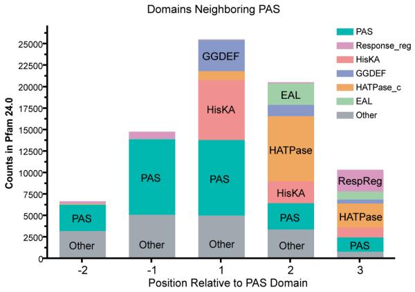
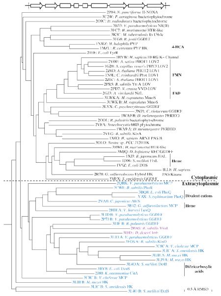
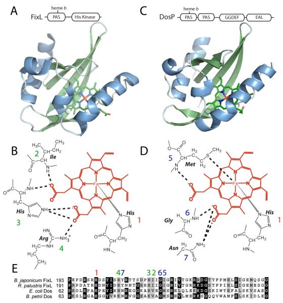
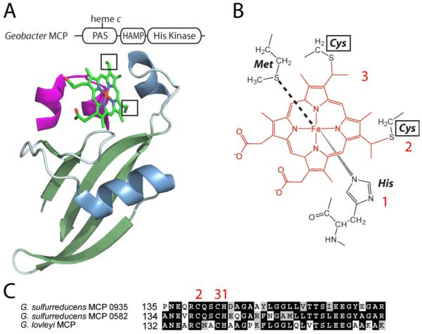
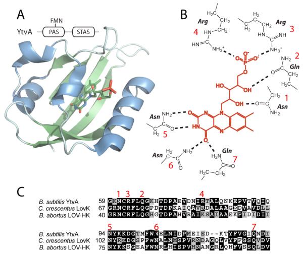
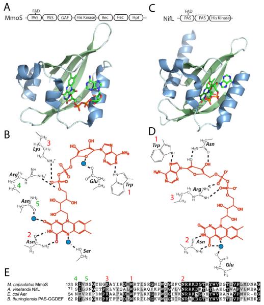
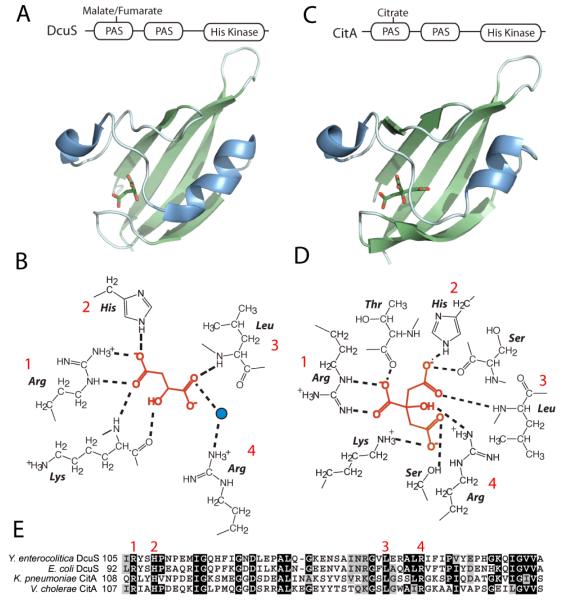
Per-Arnt-Sim (PAS) domains occur in proteins from all kingdoms of life. In the bacterial kingdom, PAS domains are commonly positioned at the amino terminus of signaling proteins such as sensor histidine kinases, cyclic-di-GMP synthases/hydrolases, and methyl-accepting chemotaxis proteins. Although these domains are highly divergent at the primary sequence level, the structures of dozens of PAS domains across a broad section of sequence space have been solved, revealing a conserved three-dimensional architecture. An all-versus-all alignment of 63 PAS structures demonstrates that the PAS domain family forms structural clades on the basis of two principal variables: (a) topological location inside or outside the plasma membrane and (b) the class of small molecule that they bind. The binding of a chemically diverse range of small-molecule metabolites is a hallmark of the PAS domain family. PAS ligand binding either functions as a primary cue to initiate a cellular signaling response or provides the domain with the capacity to respond to secondary physical or chemical signals such as gas molecules, redox potential, or photons. This review synthesizes the current state of knowledge of the structural foundations and evolution of ligand recognition and binding by PAS domains.


Figures

Figure 1
Figure 1
Predicted Domains contained in PAS Proteins. We calculated the proportions of all PAS proteins (annotated by one of the seven PAS Clan CL0183 HMMs in Pfam 24.0) containing other Pfam-annotated domains (34). For the special case of the PAS domain itself, we calculate the proportion of PAS proteins containing two or more PAS domains. Counts for domains with similar functions were combined, and the highest scoring twelve domain types are presented here. Counts for each individual domain are tabulated in Supplementary Table 1.
Figure 2
Figure 2
Domains that are neighbors of PAS, by position. For each individual PAS domain annotated in Pfam (34), we identified other annotated domains at its N- and C-terminus in the same protein. The PAS domain occupies position zero, the −1 position represents the nearest domain annotated N-terminal to the PAS domain, and the +1 position represents the nearest domain annotated C-terminal. Subsequent positions are separated from the PAS domain in question by intervening domains. A full count of all domains neighboring PAS domains by position is found in Supplementary Table 2.
Figure 3
Figure 3
Structural relatedness between PAS and PAS-like domains. PDB coordinates for representatives of what we believe to be all known PAS structures at the time of writing were trimmed to include only the region between the beginning of the first β-strand (Aβ) and the end of the final β-strand (Iβ); cofactors, ligands, and flanking sequences were discarded. The aligned region is illustrated in the secondary structure diagram, in which arrows represent β-strands and boxes represent α-helices present in most structures. We note that some sequences corresponding to the included structures are annotated as Cache domains in Genbank (8) or unrecognized by current HMM-based annotation; others are erroneously annotated as possessing a “heme pocket.” Generally, the ‘A’ chains of multi-chain files were used unless other chains were more fully resolved; for structures in which multiple PAS domains are present on a single chain, the N- and C-terminal domains are referred to as ‘A’ and ‘B’ respectively. All structures were further annotated according to their organism of origin, given name and/or predicted or known signaling output domain. Structures were oriented in PyMOL (99), then multiply aligned using the Structural Alignment of Multiple Proteins (STAMP) algorithm (96) with the MultiSeq extension of the VMD software package (30). For all structures for which reliable multiple alignment was obtained, a distance matrix of RMSD values was exported to FITCH (Phylip) for clustering by the Fitch-Margoliash method (33; 36). Black text indicates cytoplasmic localization, blue indicates extracytoplasmic localization, and purple denotes cytoplasmic PAS-like structures included in the alignment. Physiological ligands and cofactors, but not fortuitous ligands of crystallization, are indicated on the right.
Figure 4
Figure 4
Structure and ligand binding in the heme b PAS sensor domains of FixL and Dos. (A) Overall domain architecture and ribbon rendering of the B. japonicum FixL PAS domain bound to heme b (PDB ID: 1DRM). β-strands are rendered in light green, α-helices in blue. Atoms of the heme cofactor bound to the PAS domain are colored by element: carbon-green; nitrogen-blue; oxygen-red; iron-orange. (B) Drawing of protein interactions with the heme b cofactor (in red) in FixL-PAS marked with dotted lines or wedge lines. Interacting residues are numbered on the line drawing and sequence alignment with red numbers indicating conserved interactions between FixL and E. coli Dos, and green numbers indicating heme interactions that are specific to FixL-PAS. (C) Overall domain architecture and ribbon rendering of E. coli Dos PAS domain bound to heme b (PDB ID: 1S66) colored as in panel A. (D) Drawing of side chain and backbone interactions with the heme b cofactor (in red) in Dos-PAS marked with dotted lines or wedge lines. Blue numbers indicate heme interactions specific to Dos-PAS. (E) Clustal sequence alignment of FixL and Dos heme b PAS domains with the position of interacting residues numbered as described above. FixL of Rhodopseudomonas palustris (gb|ACF03217) and the DosP-like protein of Bordatella petrii (emb|CAP41703) are shown for comparison.
Figure 5
Figure 5
Structure and ligand binding in the heme c PAS sensor domains of chemoreceptor proteins. (A) Overall domain architecture and ribbon rendering of G. sulfurreducens MCP GSU0935 bound to heme c (PDB ID: 3B42). β-strands are rendered in light green, α-helices in blue. Atoms of the heme cofactor bound to the PAS domain are colored by element: carbon-green; nitrogen-blue; oxygen-red; iron-orange. (B) Drawing of protein interactions with the heme c cofactor (in red) in GSU0935-PAS marked with dotted lines or wedge lines. Interacting residues are numbered on the line drawing and sequence alignment with red numbers indicating conserved interactions between heme and MCPs of G. sulfurreducens. (C) Clustal sequence alignment of chemoreceptor heme c PAS domains with the position of interacting residues numbered as described above.
Figure 6
Figure 6
Structure and flavin binding in the class of PAS blue light photosensors known as LOV domains. (A) Overall domain architecture and ribbon rendering of B. subtilis YtvA bound to flavin mononucleotide (FMN) (PDB ID: 2PR5). β-strands are rendered in light green, α-helices in blue. Atoms of the flavin cofactor bound to the PAS domain are colored by element: carbon-green; nitrogen-blue; oxygen-red. (B) Drawing of side chain interactions with the flavin cofactor (in red) in LOV domains. Interacting residues are numbered on the line drawing and sequence alignment with red numbers indicating conserved interactions between protein and the flavin cofactor. (C) Clustal sequence alignment of YtvA and two LOV histidine kinases of C. crescentus and B. abortus, with the position of conserved interacting residues numbered as described above.
Figure 7
Figure 7
Structure and ligand binding in flavin adenine dinucleotide (FAD)-binding PAS redox sensor domains of MmoS and NifL. (A) Overall domain architecture and ribbon rendering of the M. capsulatus MmoS PAS1 domain bound to FAD (PDB ID: 3EWK). β-strands are rendered in light green, α-helices in blue. Atoms of the flavin cofactor bound to the PAS1 domain are colored by element: carbon-green; nitrogen-blue; oxygen-red. (B) Drawing of side chain and backbone interactions with the flavin cofactor (in red) in MmoS-PAS1 marked with dotted lines. Bridging water molecules are shown as blue circles. Interacting residues are numbered on the line drawing and sequence alignment with red numbers indicating conserved interactions between MmoS and A. vinelandii NifL-PAS1, and green numbers indicating flavin interactions that are specific to MmoS-PAS1. (C) Overall domain architecture and ribbon rendering of A. vinelandii NifL-PAS1 domain bound to FAD (PDB ID: 2GJ3) colored as in panel A. (D) Drawing of side chain and backbone interactions with the FAD (in red) in NifL-PAS1 marked with dotted lines. Bridging water molecules are shown as blue circles. (E) Clustal sequence alignment of MmoS and NifL PAS1 domains with the position of FAD interacting residues numbered as described above. Related PAS domains of the E. coli aerotaxis sensor, Aer (NP_417543), and B. thuringiensis PAS-GGDEF (ZP_04100494) are shown for comparison.
Figure 8
Figure 8
Structure and ligand binding in the related di-/tricarboxylate-binding PAS domains of the DcuS and CitA sensor histidine kinases. (A) Overall domain architecture and ribbon rendering of E. coli DcuS PAS1 domain bound to a malate ion (PDB ID: 3BY8). β-strands are rendered in light green, α-helices in blue. Atoms of the malate cofactor bound to the PAS1 domain are colored by element: carbon-green; oxygen-red. (B) Drawing of side chain and backbone interactions of DcuS-PAS1 with malate (in red) marked with dotted lines. Interacting residues are numbered on the line drawing and sequence alignment with red numbers indicating conserved interactions between DcuS and Klebsiella pneumoniae CitA-PAS1. (C) Overall domain architecture and ribbon rendering of K. pneumoniae CitA-PAS1 domain bound to a citrate ion (PDB ID: 1P0Z) colored as in panel A. (D) Drawing of side chain and backbone interactions with citrate (in red) in CitA-PAS1 marked with dotted lines. (E) Clustal sequence alignment of DcuS and CitA PAS1 domains with the position of ligand interacting residues numbered as described above. Related PAS domains of Yersinia enterocolitica, DcuS (YE2505), and Vibrio cholerae CitA (VC0791) are shown for comparison.
Figure 9
Figure 9
Structure and ligand binding in the dicarboxylate-binding PAS1 domain of DctB. (A) Overall domain architecture and ribbon rendering of Sinorhizobium meliloti DctB PAS1 domain bound to a succinate ion (PDB ID: 3E4O). β-strands are rendered in light green, α-helices in blue. Atoms of the succinate cofactor bound to the PAS1 domain are colored by element: carbon-green; oxygen-red. (B) Drawing of side chain and backbone interactions of DctB-PAS1 with succinate (in red) marked with dotted lines. Interacting residues are numbered on the line drawing and sequence alignment with red numbers indicating conserved interactions with DctB orthologs of A. vinelandii and Burkholderia pseudomallei. (C) Clustal sequence alignment of DctB PAS1 domains with the position of ligand interacting residues numbered as described above. Related PAS domains of A. vinelandii, DctB, and Burkholderia pseudomallei DctB are shown for comparison.
Figure 10
Figure 10
Structure and metal binding in the PAS domain of PhoQ. (A) Overall domain architecture and ribbon rendering of Salmonella typhimurium PhoQ PAS domain bound to Ca2+ ions (PDB ID: 1YAX). β-strands are rendered in light green, α-helices in blue; calcium ions colored in red. (B) Drawing of protein interactions of PhoQ-PAS with calcium cations (in red) marked with dotted lines. Ion interactions pictured are boxed in panel A. Bridging water molecules are colored as blue circles. Interacting residues are numbered on the line drawing and sequence alignment with red numbers indicating conserved interactions with PhoQ orthologs of E. coli and Yersinia pestis. (C) Clustal sequence alignment of PhoQ PAS domains with the position of ligand interacting residues numbered as described above. Related PAS domains of E. coli, PhoQ, and Y. pestis PhoQ are shown for comparison.
Figure 11
Figure 11
Structure and ligand binding in the PAS domain, photoactive yellow protein (PYP). (A) Overall domain architecture and ribbon rendering of Halorhodospira halophila PYP bound to 4-hydroxycinnamic acid (4-HCA) (PDB ID: 2PHY). β-strands are rendered in light green, α-helices in blue. Atoms of the 4-HCA cofactor bound to PYP are colored by element: carbon-green; oxygen-red. (B) Drawing of side chains of PYP that form the covalent linkage and direct polar interactions with 4-HCA (in red). Interacting residues are numbered on the line drawing and sequence alignment with red numbers indicating conserved interactions across related PYP domains of Stigmatella aurantiaca and Rhodospirillum centenum. (C) Clustal sequence alignment of PYP domains with the position of ligand interacting residues numbered as described above. Related PYPs of S. aurantiaca and R. centenum are shown for comparison.

References

    1. Akbar S, Gaidenko TA, Kang CM, O’Reilly M, Devine KM, Price CW. New family of regulators in the environmental signaling pathway which activates the general stress transcription factor sigma(B) of Bacillus subtilis. J Bacteriol. 2001;183:1329–38. - PMC - PubMed
    1. Amezcua CA, Harper SM, Rutter J, Gardner KH. Structure and interactions of PAS kinase N-terminal PAS domain: model for intramolecular kinase regulation. Structure. 2002;10:1349–61. - PubMed
    1. Anantharaman V, Koonin EV, Aravind L. Regulatory potential, phyletic distribution and evolution of ancient, intracellular small-molecule-binding domains. J Mol Biol. 2001;307:1271–92. - PubMed
    1. Anderson S, Crosson S, Moffat K. Short hydrogen bonds in photoactive yellow protein. Acta Crystallogr D Biol Crystallogr. 2004;60:1008–16. - PubMed
    1. Aravind L, Ponting CP. The GAF domain: an evolutionary link between diverse phototransducing proteins. Trends Biochem Sci. 1997;22:458–9. - PubMed

LinkOut - more resources