. 2011 Jun 10:6:28.
doi: 10.1186/1749-8104-6-28.

A conserved juxtacrine signal regulates synaptic partner recognition in Caenorhabditis elegans

Affiliations

A conserved juxtacrine signal regulates synaptic partner recognition in Caenorhabditis elegans

Joori Park et al. Neural Dev. .

Abstract

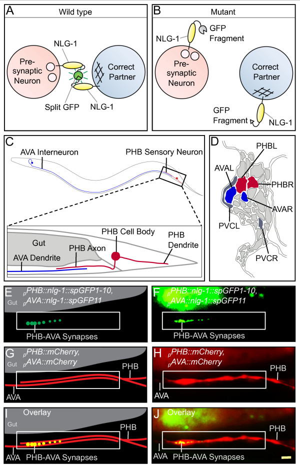
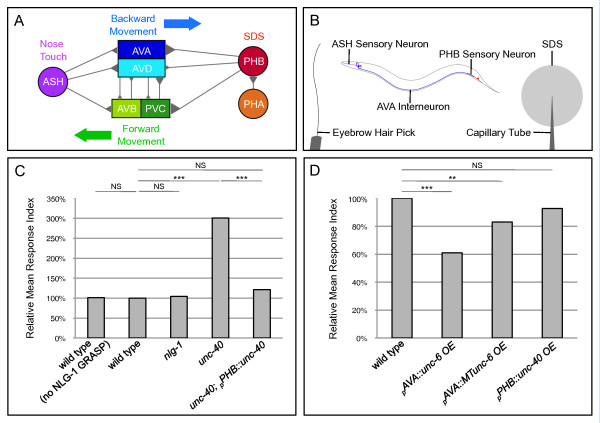
Background: An essential stage of neural development involves the assembly of neural circuits via formation of inter-neuronal connections. Early steps in neural circuit formation, including cell migration, axon guidance, and the localization of synaptic components, are well described. However, upon reaching their target region, most neurites still contact many potential partners. In order to assemble functional circuits, it is critical that within this group of cells, neurons identify and form connections only with their appropriate partners, a process we call synaptic partner recognition (SPR). To understand how SPR is mediated, we previously developed a genetically encoded fluorescent trans-synaptic marker called NLG-1 GRASP, which labels synaptic contacts between individual neurons of interest in dense cellular environments in the genetic model organism Caenorhabditis elegans.

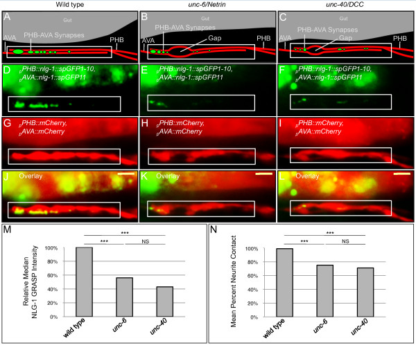
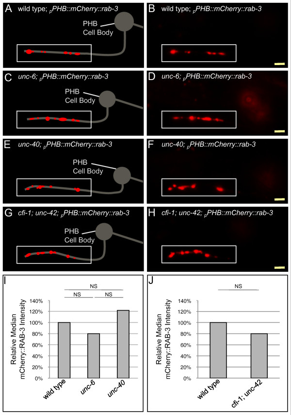
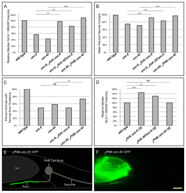
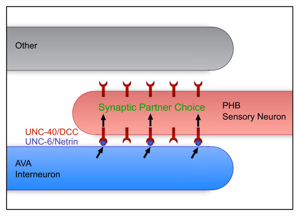
Results: Here, we describe the first use of NLG-1 GRASP technology, to identify SPR genes that function in this critical process. The NLG-1 GRASP system allows us to assess synaptogenesis between PHB sensory neurons and AVA interneurons instantly in live animals, making genetic analysis feasible. Additionally, we employ a behavioral assay to specifically test PHB sensory circuit function. Utilizing this approach, we reveal a new role for the secreted UNC-6/Netrin ligand and its transmembrane receptor UNC-40/Deleted in colorectal cancer (DCC) in SPR. Synapses between PHB and AVA are severely reduced in unc-6 and unc-40 animals despite normal axon guidance and subcellular localization of synaptic components. Additionally, behavioral defects indicate a complete disruption of PHB circuit function in unc-40 mutants. Our data indicate that UNC-40 and UNC-6 function in PHB and AVA, respectively, to specify SPR. Strikingly, overexpression of UNC-6 in postsynaptic neurons is sufficient to promote increased PHB-AVA synaptogenesis and to potentiate the behavioral response beyond wild-type levels. Furthermore, an artificially membrane-tethered UNC-6 expressed in the postsynaptic neurons promotes SPR, consistent with a short-range signal between adjacent synaptic partners.

Conclusions: These results indicate that the conserved UNC-6/Netrin-UNC-40/DCC ligand-receptor pair has a previously unknown function, acting in a juxtacrine manner to specify recognition of individual postsynaptic neurons. Furthermore, they illustrate the potential of this new approach, combining NLG-1 GRASP and behavioral analysis, in gene discovery and characterization.


Figures

Figure 1
Figure 1
NLG-1 GRASP labels specific synapses between PHB sensory neurons and AVA interneurons. (A) Schematic diagram of NLG-1 GRASP, which uses split GFP to label synapses formed correctly between pre- and postsynaptic neurons. (B) If a neurite fails to identify the correct partner, NLG-1 GRASP will not reconstitute. (C) The PHB sensory neurons form synapses with the AVA interneurons in the preanal ganglion (one neuron from each pair is depicted for simplicity). (D) Schematic of an electron micrograph representing the preanal ganglion in cross section from a gravid adult, displaying PHB neurons (red) and their primary postsynaptic partners AVA (blue) and PVC (gray) (adapted from [20]). (E,F) Schematic (E) and micrograph (F) of the NLG-1 GRASP marker labeling synapses between PHB and AVA neurons. The signal in the dorsal region is gut autofluorescence. (G,H) Schematic (G) and micrograph (H) of mCherry-labeled PHB and AVA neurites in contact. (I,J) Schematic (I) and micrograph (J) of merged image. White boxes indicate the region of neurite overlap. Yellow scale bar: 2 μm.
Figure 2
Figure 2
unc-6 and unc-40 mutants display defective SPR. (A) Schematic and (D,G,J) micrographs of a wild-type animal. (D) The NLG-1 GRASP signal indicates synapses between PHB and AVA neurons. (G) mCherry labels PHB and AVA neurites and shows nearly complete contact. (J) Merged image. (B) Schematic and (E,H,K) micrographs of an unc-6 mutant animal. (E) The NLG-1 GRASP signal is severely reduced. (H) Minor defects in neurite contact are visualized as small gaps between mCherry-labeled neurites. (K) Merge. (C) Schematic and (F,I,L) micrographs of an unc-40 mutant. (F) The reduced NLG-1 GRASP signal is similar to that in unc-6 animals. (I) Minor defects in PHB and AVA neurite contact are also similar. (L) Merged image. Yellow scale bar: 2 μm. (M) Quantification of reduction in NLG-1 GRASP fluorescence in unc-6 and unc-40 mutants using NIH ImageJ. Wild-type n = 94, unc-6 n = 87, and unc-40 n = 85 animals. ***P < 0.001, NS, not significant, u-test. P-values were adjusted for multiple comparisons using the Hochberg method. (N) Quantification of contact defects between PHB and AVA neurites using NIH ImageJ. Wild-type n = 94, unc-6 n = 87, and unc-40 n = 85 animals. ***P < 0.001, NS, not significant, t-test. P-values were adjusted for multiple comparisons using the Hochberg method.
Figure 3
Figure 3
Presynaptic components localize to the correct subcellular compartment in unc-6 and unc-40 mutant animals. (A,B) Wild type, (C,D) unc-6, (E,F) unc-40, and (G,H) cfi-1; unc-42 animals are labeled with the presynaptic vesicle marker mCherry::rab-3 expressed in PHB neurons. Presynaptic specializations are unaltered in unc-6, unc-40, and cfi-1; unc-42 mutant animals and localize to the distal region of the PHB axon within the preanal ganglion, where PHB synapses normally form (boxed in white). Yellow scale bar: 2 μm. (I) Quantification of mCherry::RAB-3 fluorescence intensity using NIH ImageJ indicates no significant difference among wild-type, unc-6 and unc-40 animals. Wild-type n = 32, unc-6 n = 47, and unc-40 n = 36 animals. NS, not significant, Kruskal-Wallis test. (J) Quantification of mCherry::RAB-3 fluorescence intensity using NIH ImageJ indicates no significant different among wild-type and cfi-1; unc-42 animals. Wild-type n = 45, and cfi-1; unc-42 n = 45 animals. NS, not significant, u-test.
Figure 4
Figure 4
Expression of UNC-6 in AVA or UNC-40 in PHB is sufficient for SPR. (A) Expression of pAVA::unc-6 in unc-6 mutants and pPHB::unc-40 in unc-40 mutants restores NLG-1 GRASP fluorescence, labeling PHB-AVA synapses. NLG-1 GRASP fluorescence is also significantly rescued in unc-6 mutants by expressing a membrane-tethered unc-6 in AVA (pAVA::MTunc-6). ***P < 0.001, **P < 0.01, u-test. (B) Neurite contact between PHB and AVA is significantly rescued by expression of pAVA::unc-6 or pAVA::MTunc-6 in unc-6 mutants and pPHB::unc-40 in unc-40 mutants. ***P < 0.001, **P < 0.01, t-test. (A,B) Two lines were examined for each transgene; in all cases, both lines gave similar results. Wild-type n = 254, unc-6 n = 87, unc-6; pAVA::unc-6 n = 70, unc-6; pAVA::MTunc-6 n = 84, unc-40 n = 85, and unc-40; pPHB::unc-40 n = 79 animals. (C) PHB axon guidance to the preanal ganglion is restored by expression of pPHB::unc-40 in unc-40 mutants but not by expression of pAVA::unc-6 in unc-6 mutants. Wild-type n = 94, unc-6 n = 180, unc-40 n = 147, unc-6; pAVA::unc-6 n = 144, and unc-40; pPHB::unc-40 n = 108 animals. **P < 0.01, NS, not significant, χ2 goodness-of-fit test. (D) Overexpression of pAVA::unc-6 and pAVA::MTunc-6 in wild-type animals increases NLG-1 GRASP fluorescence intensity, indicating an increase in PHB-AVA synapses, while overexpressing pPHB::unc-40 does not, indicating that the UNC-6 signal is limiting. Two lines were examined for each transgene with the exception of pPHB::unc-40 overexpression, where three lines were examined; in all cases, all lines gave similar results. Wild-type n = 165, pAVA::unc-6OE n = 82, pAVA::MTunc-6OE n = 79, and pPHB::unc-40OE n = 119 animals. ***P < 0.001, **P < 0.01, NS, not significant, u-test. (A-D) P-values were adjusted for multiple comparisons using the Hochberg method. (E) Schematic and (F) micrograph of UNC-40 subcellular localization. UNC-40::GFP is localized to the region of contact with AVA within the PHB axon, but is excluded from the commissure and dendrite. Yellow scale bar: 5 μm.
Figure 5
Figure 5
A PHB circuit-specific behavioral assay indicates that UNC-40 and UNC-6 mediate formation of functional synapses. (A) Neural circuit diagram summarizing synaptic contacts between neurons in PHB and ASH sensory circuits. (B) Behavioral assay outline. Function of the PHB circuit is tested by stimulating ASH neurons. ASH-AVA synapses control backward movement induced by a nose touch with an eyebrow hair pick, and PHB-AVA synapses control termination of backwards movement in response to the noxious chemical SDS. (C) Neither the NLG-1 GRASP marker nor mutations in nlg-1 affect SDS sensitivity. However, unc-40 mutants fail to respond to SDS, indicating complete loss of PHB-circuit function. Cell-autonomous expression of unc-40 in PHB neurons rescues the behavioral defect. unc-6 animals are too uncoordinated to be examined with this assay. Two lines were examined for each transgene; in all cases, both lines gave similar results. Wild-type n = 200, wild-type (no NLG-1 GRASP) n = 40, nlg-1 n = 40, unc-40 n = 40, unc-40; pPHB::unc-40 n = 80 animals. ***P < 0.001, NS, not significant, t-test. P-values were adjusted for multiple comparisons using the Hochberg method. (D) Overexpression of unc-6 and MTunc-6 in AVA neurons potentiates the behavioral response while overexpression of unc-40 in PHB does not, indicating that UNC-6 is limiting. Two lines were examined for each transgene with the exception of pPHB::unc-40 overexpression, where three lines were examined; in all cases, all lines gave similar results. Wild-type n = 200, pAVA::unc-6 overexpression n = 80, pAVA::MTunc-6 overexpression n = 80, and pPHB::unc-40 overexpression n = 80 animals. ***P < 0.001, **P < 0.01, NS, not significant, t-test. P-values were adjusted for multiple comparisons using the Hochberg method.
Figure 6
Figure 6
Limiting amounts of UNC-6/Netrin promote SPR through a juxtacrine signal from postsynaptic to presynaptic neurons. In this model, UNC-6 secreted from AVA interneurons binds UNC-40 expressed in PHB neurons to direct SPR. Limiting amounts of UNC-6 sequestered near the AVA membrane bind a subset of the available UNC-40 receptors in PHB, inducing a recognition event that results in correct adhesion and synaptogenesis between the two neurons.

References

    1. Shen K, Scheiffele P. Genetics and cell biology of building specific synapse connectivity. Annu Rev Neurosci. 2010;33:473–507. doi: 10.1146/annurev.neuro.051508.135302. - DOI - PMC - PubMed
    1. Hamos JE, Van Horn SC, Raczkowski D, Sherman SM. Synaptic circuits involving an individual retinogeniculate axon in the cat. J Comp Neurol. 1987;259:165–192. doi: 10.1002/cne.902590202. - DOI - PubMed
    1. White JG, Southgate E, Thomson JN, Brenner S. The structure of the nervous system of the nematode C. elegans. Philos Trans R Soc Lond B Biol Sci. 1986;314:1–340. doi: 10.1098/rstb.1986.0056. - DOI - PubMed
    1. Moore SW, Tessier-Lavigne M, Kennedy TE. Netrins and their receptors. Adv Exp Med Biol. 2007;621:17–31. doi: 10.1007/978-0-387-76715-4_2. - DOI - PubMed
    1. Dickson BJ. Molecular mechanisms of axon guidance. Science. 2002;298:1959–1964. doi: 10.1126/science.1072165. - DOI - PubMed

Publication types

MeSH terms