. 2011 Jun 10;12(1):77.
doi: 10.1186/1465-9921-12-77.

CD39+ regulatory T cells suppress generation and differentiation of Th17 cells in human malignant pleural effusion via a LAP-dependent mechanism

Affiliations

CD39+ regulatory T cells suppress generation and differentiation of Th17 cells in human malignant pleural effusion via a LAP-dependent mechanism

Zhi-Jian Ye et al. Respir Res. .

Abstract

Background: Both regulatory T cells (Tregs) and T helper IL-17-producing cells (Th17 cells) have been found to be involved in human malignancies, however, the possible implication of Tregs in regulating generation and differentiation of Th17 cells in malignant pleural effusion remains to be elucidated.

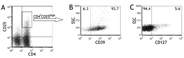
Methods: The numbers of both CD39(+)Tregs and Th17 cells in malignant pleural effusion and peripheral blood from patients with lung cancer were determined by flow cytometry. The regulation and mechanism of Tregs on generation and differentiation of Th17 cells were explored.

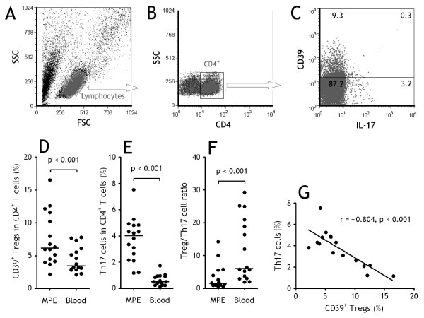
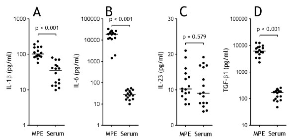
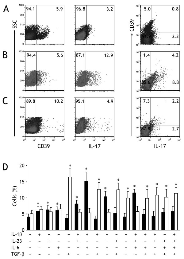
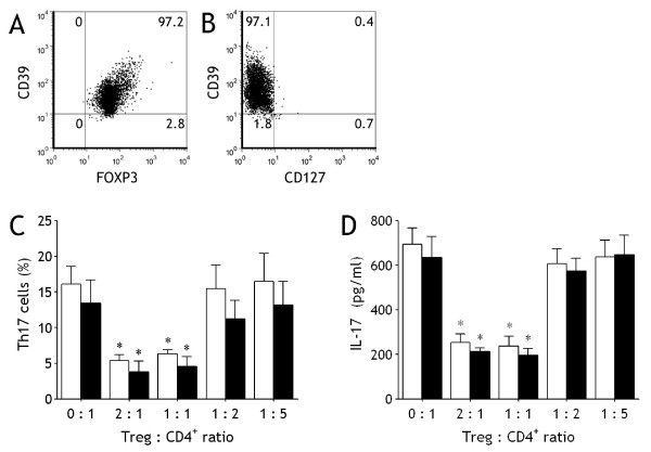
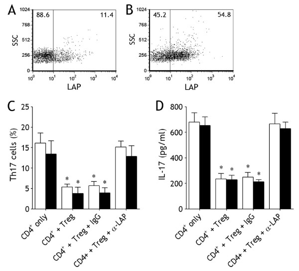
Results: Both CD39(+)Tregs and Th17 cells were increased in malignant pleural effusion when compared with blood, and the numbers of CD39(+)Tregs were correlated negatively with those of Th17 cells. It was also noted that high levels of IL-1β, IL-6, and TGF-β1 could be observed in malignant pleural effusion when compared the corresponding serum, and that pleural CD39(+)Tregs could express latency-associated peptide on their surface. When naïve CD4(+) T cells were cocultured with CD39(+)Tregs, Th17 cell numbers decreased as CD39(+)Treg numbers increased, addition of the anti-latency-associated peptide mAb to the coculture reverted the inhibitory effect exerted by CD39(+)Tregs.

Conclusions: Therefore, the above results indicate that CD39(+)Tregs inhibit generation and differentiation of Th17 cells via a latency-associated peptide-dependent mechanism.


Figures

Figure 1
Figure 1
Both CD39+ regulatory T (Tregs) cells and Th17 cells increased in malignant pleural effusion (MPE). (A) Lymphocytes were identified based on their characteristic properties shown in the forward scatter (FSC) and sideward scatter (SSC). (B) A representative gating was set for CD4+ T cells from pleural lymphocytes. (C) A representative dot plots showing expression of CD39 and IL-17 in pleural CD4+ T cells. Comparisons of percentages of CD39+Tregs (D), Th17 cells (E), and ratios of CD39+Tregs/Th17 cells (F) in MPE and blood from patients with lung cancer (n = 16). The percentages of CD39+Tregs and Th17 cells were determined by flow cytometry. Horizontal bars indicate medians. Comparison was made using a Wilcoxon signed-rank test. (G) The percentages of Th17 cells correlated with CD39+Tregs cells in MPE. Correlations were determined by Spearman's rank correlation coefficients.
Figure 2
Figure 2
CD39 and CD127 expressed on CD4+CD25high T cells. The subset of pleural CD4+CD25high T cells (A) was identified by flow cytometry for determining the surface expression of CD39 (B) and CD127 (C), data for one representative donor of 16 tested are shown.
Figure 3
Figure 3
Proinflammatory cytokines increased in malignant pleural effusion (MPE). Comparisons of concentrations of IL-1β (A), IL-6 (B), IL-23 (C), and TGF-β1 (D) in both MPE and sera from patients with lung cancer (n = 16). The cytokines were determined by ELISA, and comparisons of cytokine concentrations were made using a Wilcoxon signed-rank test.
Figure 4
Figure 4
Generation and differentiation of human CD39+Tregs and Th17 cells from malignant pleural effusion regulated by different cytokines. (A) The representative dot plots of freshly isolated naïve CD4+ T cells from malignant pleural effusion were determined for expression of CD39 and IL-17 by flow cytometry. (B) The representative dot plots of CD39+Tregs and Th17 cells detected in naïve CD4+ T cells after culturing in presence both IL-1β and IL-6. (C) The representative dot plots of CD39+Tregs and Th17 cells detected in naïve CD4+ T cells after culturing in presence of TGF-β1. (D) The mean ± SEM of CD39+Tregs (open bars) and Th17 cells (closed bars) detected in naïve CD4+ T cells from 5 independent experiments. The purified naïve CD4+ T cells were stimulated with plate-bound anti-CD3 and soluble anti-CD28 mAbs in the presence of the indicated cytokines, either alone or in various combinations for 7 d. * p < 0.01 compared with their corresponding controls with no cytokines.
Figure 5
Figure 5
CD39+Tregs inhibit generation and differentiation of Th17 cells. The representative dot plots showing isolated pleural CD39+CD4+CD25high T cells are almost CD39 positive (A) and CD127 negative (B). Naïve CD4+ T cells isolated from malignant pleural effusion (open bars) and blood (closed bars) were cultured in the conditions described in Figure 4 with indicated ratio of CD39+Tregs, Th17 cell numbers were determined by flow cytometry (C) and IL-17 concentrations in the cultured supernatants were determined by ELISA (D). The results are reported as mean ± SEM from 5 independent experiments. * p < 0.01 compared with naïve CD4+ T cells without CD39+Tregs.
Figure 6
Figure 6
LAP mediates Treg-induced inhibition of Th17 cells. Freshly purified pleural CD39+Tregs (A) and cultured CD39+Tregs (B) were analyzed by flow cytometry for determining the surface expression of LAP, data for one representative donor of 5 tested are shown. Naïve CD4+ T cells isolated from malignant pleural effusion (open bars) and blood (closed bars) were cultured CD39+Tregs (ratio, 1 : 1), an anti-LAP mAb or isotype control IgG was added into the coculture, Th17 cell numbers were determined by flow cytometry (C) and IL-17 concentrations in the cultured supernatants were determined by ELISA (D). The results are reported as mean ± SEM from 5 independent experiments. * p < 0.01 compared with isotype control.

References

    1. Light RW. Clinical practice. Pleural effusion. N Engl J Med. 2002;346:1971–1977. doi: 10.1056/NEJMcp010731. - DOI - PubMed
    1. Lucivero G, Pierucci G, Bonomo L. Lymphocyte subsets in peripheral blood and pleural fluid. Eur Respir J. 1988;1:337–340. - PubMed
    1. Ikeda H, Chamoto K, Tsuji T, Suzuki Y, Wakita D, Takeshima T, Nishimura T. The critical role of type-1 innate and acquired immunity in tumor immunotherapy. Cancer Sci. 2004;95:697–703. doi: 10.1111/j.1349-7006.2004.tb03248.x. - DOI - PMC - PubMed
    1. Atanackovic D, Block A, de WA, Faltz C, Hossfeld DK, Hegewisch-Becker S. Characterization of effusion-infiltrating T cells: benign versus malignant effusions. Clin Cancer Res. 2004;10:2600–2608. doi: 10.1158/1078-0432.CCR-03-0239. - DOI - PubMed
    1. Hori S, Nomura T, Sakaguchi S. Control of regulatory T cell development by the transcription factor Foxp3. Science. 2003;299:1057–1061. doi: 10.1126/science.1079490. - DOI - PubMed

Publication types

MeSH terms