. 2011 Jun 10:11:19.
doi: 10.1186/1475-2867-11-19.

Vitamin K3 and vitamin C alone or in combination induced apoptosis in leukemia cells by a similar oxidative stress signalling mechanism

Affiliations

Vitamin K3 and vitamin C alone or in combination induced apoptosis in leukemia cells by a similar oxidative stress signalling mechanism

Angelica R Bonilla-Porras et al. Cancer Cell Int. .

Abstract

Background: Secondary therapy-related acute lymphoblastic leukemia might emerge following chemotherapy and/or radiotherapy for primary malignancies. Therefore, other alternatives should be pursued to treat leukemia.

Results: It is shown that vitamin K3- or vitamin C- induced apoptosis in leukemia cells by oxidative stress mechanism involving superoxide anion radical and hydrogen peroxide generation, activation of NF-κB, p53, c-Jun, protease caspase-3 activation and mitochondria depolarization leading to nuclei fragmentation. Cell death was more prominent when Jurkat and K562 cells are exposed to VC and VK3 in a ratio 1000:1 (10 mM: 10 μM) or 100:1 (300 μM: 3 μM), respectively.

Conclusion: We provide for the first time in vitro evidence supporting a causative role for oxidative stress in VK3- and VC-induced apoptosis in Jurkat and K562 cells in a domino-like mechanism. Altogether these data suggest that VK3 and VC should be useful in the treatment of leukemia.


Figures

Figure 1
Figure 1
Vitamin K3 (VK3) and vitamin C (VC) produce reactive oxygen species, mitochondrial depolarization and nuclear morphology indicative of apoptosis in lymphocyte and leukemia cells. Lymphocyte, Jurkat and K562 were incubated with increasing concentration of VK3 (A) and VC (B) for 24 h. Nuclear morphological changes was evaluated using AO/EB staining. Inset: Representative fluorescent photomicrography shows cell shrinkage and rounding, chromatin condensation (arrows), nuclei fragmentation (arrowheads) indicative of apoptosis and necrotic cells (asterisk) in K562 cells treated with (35 μM) VK3 (A) and (1 mM) VC (B). The ANOVA showed significantly differences among the three cells groups, p < 0.0001; post-hoc comparison showed significantly increase in cell death morphology in the leukemia cells on each VK3 and VC concentration versus lymphocytes. Jurkat cells were incubated with (10 μM) VK3 (C) and (10 mM) VC (D) for 24 h. Cells were evaluated for O2.-/H2O2 production, Δψm and nuclear morphological changes indicative of apoptosis. NBT+ stained blue-purple precipitate cells, DCF+ green fluorescent cells, DiOC6(3)high/low+ green fluorescent cells and apoptotic nuclei percentage is expressed as mean of percentage (%) ± S.D. from three independent experiments. ANOVA test for each condition showed differences among groups p < 0.0001. The post-hoc comparison showed increase in number of DCF, AO/EB and NBT+ cells, whereas the number of DiOC6(3)+ cells decreased in a concentration-dependent fashion. One-way ANOVA analysis with Bonferroni post-hoc analysis was performed. A p-value of ap < 0.05 and bp < 0.001 Jurkat versus K562 or cp < 0.05 and dp < 0.001 versus lymphocytes (A, B) or control (C, D) was considered significant.
Figure 2
Figure 2
Vitamin K3 (VK3) generates anion superoxide radicals (O2.-), hydrogen peroxide (H2O2), mitochondrial depolarization and apoptotic morphology in Jurkat cells. Jurkat cells (1 × 10⁶ cells/mL) were incubated with (10 μM) VK3 at different interval as indicated below. (A) Representative light photomicrography showing NBT+ stained blue-purple precipitate cells (i.e. formazan, arrows) and NBT- stained cells (i.e. translucent cells, arrowheads) indicative of positive and negative (O2.-) production, respectively from Jurkat cells incubated with VK3 for 3 h at 37 °C. Insets: Magnification of four NBT+ cells showing cytoplasmic blue-purple precipitate from Jurkat cells incubated with VK3 for 24 h at 37 °C. (B) Representative fluorescent photomicrography (ex. 450-490 nm, em. 515 nm) illustrating DCF+ (green bright light, arrowheads) and DCF- (dark round-shape, asterisks) fluorescent stained cells indicative of positive and negative H2O2 production, respectively. (C) Representative fluorescent photomicrography (ex. 450-490 nm, em. 515 nm) illustrating DiOC6(3)+ green fluorescent (arrowheads) stained cells indicative of high-polarized and low-polarized mitochondria and DiOC6(3)- (non-fluorescent, asterisks) stained cells indicative of depolarized mitochondria. (D) Representative fluorescent photomicrography (ex. 450-490 nm, em. 515 nm) showing typical nuclear apoptotic morphology such as highly condensed chromatin (asterisks) and nuclear fragmentation (arrowheads) from Jurkat cells treated with (10 μM) VK3. Magnification A: 1000x (inset 1200x); B: 600x; C: 800x; D: 1000x.
Figure 3
Figure 3
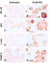
Vitamin K3 (VK3) alone induces simultaneous activation of the transcription factor NF-κB, p53, c-Jun and protease caspase-3 in Jurkat cells. Jurkat cells were left untreated (A, C, E, G) or exposed to (10 μM) VK3 (B, D, F, H), at 37 °C for 24 h. After this time of incubation, cells were stained with anti-NF-κB-p65 (A, B), anti-p53 (C, D), anti-c-Jun (E, F) and anti-caspase-3 (G, H) antibodies according to procedure described in Materials and Methods. Notice that NF-κB, p53, c-Jun and caspase-3 (CASP-3) positive-nuclei (dark brown color) reflect their nuclear translocation/activation and appear to correlate with the apoptotic nuclear morphology, i.e. condensed/fragmented nuclei compared with untreated cells (A, C, E, G) or cytoplasmic activation (brown color). Magnification 660x (A-H). Inset Magnification (B, 2000x; D:1100x).
Figure 4
Figure 4
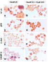
Vitamin C (VC) alone or in combination with vitamin K3 (VK3: VC) induce simultaneous activation of the transcription factor NF-κB, p53, c-Jun and protease caspase-3 in Jurkat cells. Jurkat cells were exposed to (10 mM) VC (A, C, E, G) and (10 μM) VK3 plus (10 mM) VC (B, D, F, H) for 24 h. After this time of incubation, cells were stained with anti-NF-κB-p65 (A, B), anti-p53 (C, D), anti-c-Jun (E, F) and anti-caspase-3 (G, H) antibodies according to procedure described in Materials and Methods. Notice that NF-κB, p53, c-Jun and caspase-3 (CASP-3) positive-nuclei (dark brown color) reflect their nuclear translocation/activation and appear to correlate with the apoptotic nuclear morphology, i.e. condensed/fragmented nuclei compared with untreated cells (Figure 3 A, C, E, G) or cytoplasmic activation (brown color). Magnification 660x (A-H). Inset Magnification (A: 2000x).

References

    1. Chen W, Wang E, Lu Y, Gaal KK, Huang Q. Therapy-related acute lymphoblastic leukemia without 11q23 abnormality: report of six cases and a literature review. Am J Clin Pathol. 2010;133(1):75–82. doi: 10.1309/AJCPYWC6AQC7BAVJ. - DOI - PubMed
    1. Gregory CD, Pound JD. Cell death in the neighbourhood: direct microenvironmental effects of apoptosis in normal and neoplastic tissues. J Pathol. 2011;223(2):177–194. - PubMed
    1. Baran I, Ganea C, Scordino A, Musumeci F, Barresi V, Tudisco S, Privitera S, Grasso R, Condorelli DF, Ursu I, Baran V, Katona E, Mocanu MM, Gulino M, Ungureanu R, Surcel M, Ursaciuc C. Effects of menadione, hydrogen peroxide, and quercetin on apoptosis and delayed luminescence of human leukemia Jurkat T-cells. Cell Biochem Biophys. 2010;58(3):169–179. doi: 10.1007/s12013-010-9104-1. - DOI - PubMed
    1. Lee W-J. The prospects of vitamin C in cancer therapy. Immune network. 2009;9:147–152. doi: 10.4110/in.2009.9.5.147. - DOI - PMC - PubMed
    1. Lamson DW, Gu YH, Plaza SM, Brignall MS, Brinton CA, Sadlon AE. The vitamin C: vitamin K3 system - enhancers and inhibitors of the anticancer effect. Altern Med Rev. 2010;15(4):345–351. - PubMed