Review
. 2011 Jun 10;145(6):835-50.
doi: 10.1016/j.cell.2011.05.019.

Chromatin connections to pluripotency and cellular reprogramming

Affiliations
Review

Chromatin connections to pluripotency and cellular reprogramming

Stuart H Orkin et al. Cell. .

Abstract

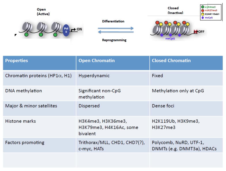
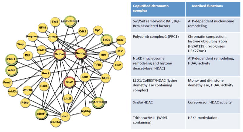
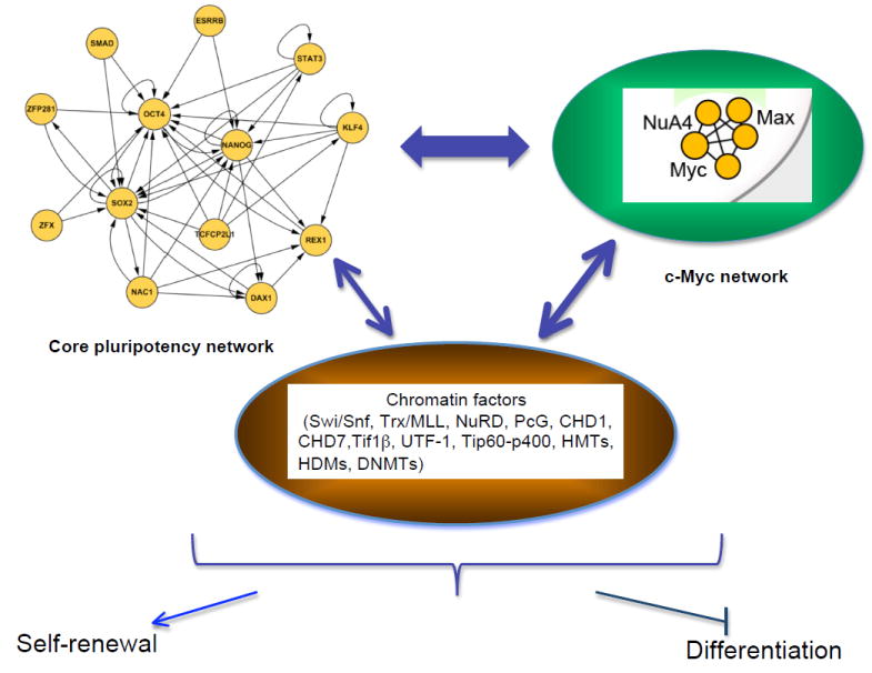
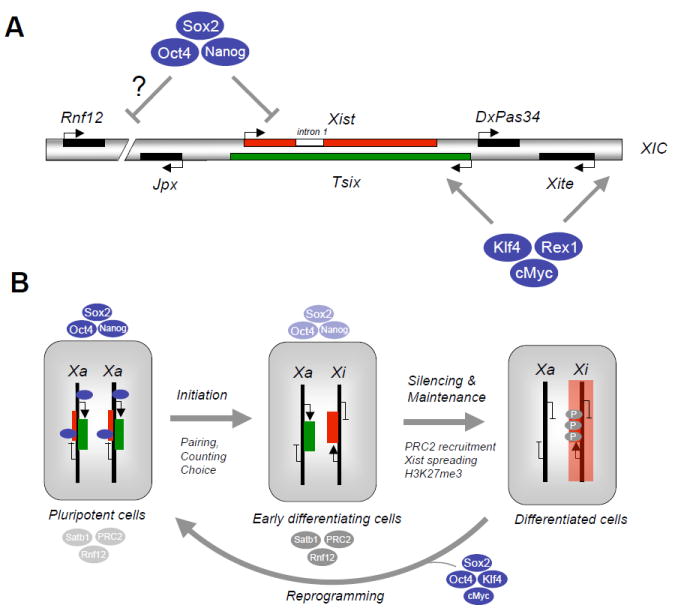
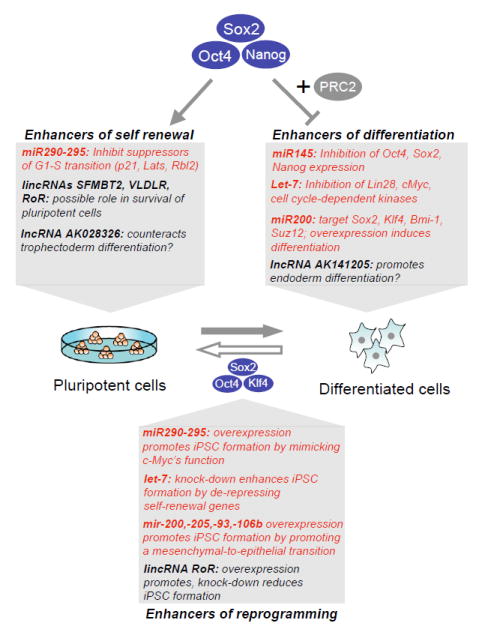
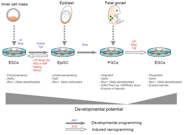
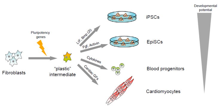
The pluripotent state of embryonic stem cells (ESCs) provides a unique perspective on regulatory programs that govern self-renewal and differentiation and somatic cell reprogramming. Here, we review the highly connected protein and transcriptional networks that maintain pluripotency and how they are intertwined with factors that affect chromatin structure and function. The complex interrelationships between pluripotency and chromatin factors are illustrated by X chromosome inactivation, regulatory control by noncoding RNAs, and environmental influences on cell states. Manipulation of cell state through the process of transdifferentiation suggests that environmental cues may direct transcriptional programs as cells enter a transiently "plastic" state during reprogramming.


Figures

Figure 1
Figure 1. Properties of open and closed chromatin
For details, (see Gaspar-Maia et al., 2011).
Figure 2
Figure 2. Protein interaction network supporting pluripotency and connections to chromatin complexes
Protein-protein interactions derived from microsequencing of protein complexes purified from ESCs are shown on the left. The triad of pluripotency factors, Oct4, Nanog, and Sox2, are circled in red. Components of chromatin remodeling or modifying complexes are highlighted in green circles. On the right, several of the protein complexes associated with the pluripotency protein network are listed with their associated functional activities.
Figure 3
Figure 3. Transcriptional regulatory interactions in ESCs
The core network depicted on the upper right illustrates target gene relationships. The c-myc network to the upper left represents the sub-network defined by common target genes (see Kim et al, 2010). The factors in the core and c-myc regulatory networks cross-regulate each other, and regulate, and are regulated by, chromatin factor components illustrated in the center. The output of these complex regulatory interactions is maintenance of self-renewal and blocking of lineage-specific differentiation.
Figure 4
Figure 4. Pluripotency factors and X chromosome inactivation
(A) Schematic depiction of X Inactivation Center (XIC) on the X chromosome with positions of non-coding RNAs Xist, Tsix, DxPas34, Xite, Jpx and the dose-dependent Xist activator Rnf12 as indicated. In undifferentiated female embryonic stem cells (ESCs), Xist (intron 1) and possibly Rnf12 are occupied and transcriptionally suppressed by Oct4, Sox2 and Nanog while Xite and Tsix are bound and transcriptionally activated by Klf4, Rex1 and cMyc. (B) In female ESCs, Xist is silenced while Tsix is activated by the pluripotency factors shown in (A). Upon differentiation, X chromosome inactivation ensues through a multistep process that involves initiation (pairing and counting of X chromosomes to choose the future inactive and active X, Xi and Xa), silencing and maintenance of the silenced X. The initiation and onset of silencing are tightly linked with the downregulation of pluripotency factors and the concomitant upregulation of chromatin regulators that mediate XCI, such as PRC2 (recruited by Xist RNA to mediate spreading of silencing along the entire X), and Satb1 (organization of active chromatin into loops). Silencing of the inactive X further results in H3K27 trimethylation by PRC2. Introduction of Oct4, Sox2, Klf4 and cMyc into differentiated cells gives rise to induced pluripotent stem cells, which is accompanied by X chromosome reactivation in mouse.
Figure 5
Figure 5. Non-coding RNAs modulate ESC self renewal and differentition as well as cellular reprogramming
Shown are examples of microRNAs (in red) and long non-coding (lnc)RNAs (in black) that are occupied and either activated by Oct4, Sox2 and Nanog, or silenced by the same factors in combination with the PRC2 repressor complex in pluripotent cells, and their roles in self renewal and differentiation. Manipulation of several non-coding RNAs in the context of induced pluripotent stem cell (iPSC) formation has been shown to enhance cellular reprogramming. Note that some miRNAs, such as members of the miR-200 family, seem to directly target PRC1 and PRC2 components, such as Bmi-1 and Suz12, respectively.
Figure 6
Figure 6. Examples of culture-induced epigenetic and developmental changes in pluripotent mouse cells
Embryonic stem cells (ESCs), derived from the inner cell mass of blastocysts, are maintained in an undifferentiated state in the presence of LIF and Bmp. Exchange of LIF and Bmp with Fgf and activin induces their differentiation into epiblast stem cells (EpiSCs), which are normally derived from the epiblast of postimplantation embryos and have limited differentiation potential. The ESC-to-EpiSC transition is accompanied by characteristic epigenetic changes, such as X inactivation and methylation silencing of Rex1 and Stella genes, which can be reversed by replating cells in LIF/Bmp or 2i, or upon overexpression of Klf2, Klf4, Nanog, or Nr5a2. When exposed to Bmp, EpiSCs continuously give rise to unipotent primordial germ cells (PGCs) that undergo genome-wide epigenetic remodeling, X reactivation and erasure of genomic imprinting. In the presence of LIF, Bmp and Fgf, these PGCs undergo dedifferentiation into pluripotent embryonic germ cells (EGCs).
Figure 7
Figure 7. Proposed synergism between pluripotency gene expression and growth factors in changing cellular identity
Introduction of individual or combinations of pluripotency genes into fibroblasts may generate a hypothetical “plastic” intermediate that is amenable to further reprogramming into induced pluripotent stem cells (iPSCs) or epiblast stem cells (EpiSCs) when exposed to LIF/Bmp (2i) or Fgf/activin, respectively. Alternatively, such intermediate cells may be converted directly into blood progenitors or cardiomyocytes when exposed to hematopoeitic cytokines or cardiac growth factors, respectively. Note that the developmental potency of resultant cells appears to depend on the provided growth conditions.

References

    1. Agarwal S, Loh YH, McLoughlin EM, Huang J, Park IH, Miller JD, Huo H, Okuka M, Dos Reis RM, Loewer S, et al. Telomere elongation in induced pluripotent stem cells from dyskeratosis congenita patients. Nature. 2010;464:292–296. - PMC - PubMed
    1. Agrelo R, Souabni A, Novatchkova M, Haslinger C, Leeb M, Komnenovic V, Kishimoto H, Gresh L, Kohwi-Shigematsu T, Kenner L, et al. SATB1 defines the developmental context for gene silencing by Xist in lymphoma and embryonic cells. Dev Cell. 2009;16:507–516. - PMC - PubMed
    1. Ancelin K, Lange UC, Hajkova P, Schneider R, Bannister AJ, Kouzarides T, Surani MA. Blimp1 associates with Prmt5 and directs histone arginine methylation in mouse germ cells. Nat Cell Biol. 2006;8:623–630. - PubMed
    1. Ang Y-S, Tsai S-Y, Lee D-F, Monk J, Su J, Ratnakumar K, Ding J, Ge Y, Darr H, Chang B, et al. Wdr5 mediates self-renewal and reprogramming via the embryonic stem cell core transcriptional network. Cell. 2011 in press. - PMC - PubMed
    1. Bantignies F, Roure V, Comet I, Leblanc B, Schuettengruber B, Bonnet J, Tixler V, Mas A, Cavalli G. Polycomb-dependent regulatoryu contacts between distant Hox loci in Drosophila. Cell. 2011;144:214–226. - PubMed

Publication types

LinkOut - more resources