. 2011 Jun 10;145(6):956-68.
doi: 10.1016/j.cell.2011.05.003.

Interlocked feedforward loops control cell-type-specific Rhodopsin expression in the Drosophila eye

Affiliations

Interlocked feedforward loops control cell-type-specific Rhodopsin expression in the Drosophila eye

Robert J Johnston Jr et al. Cell. .

Abstract

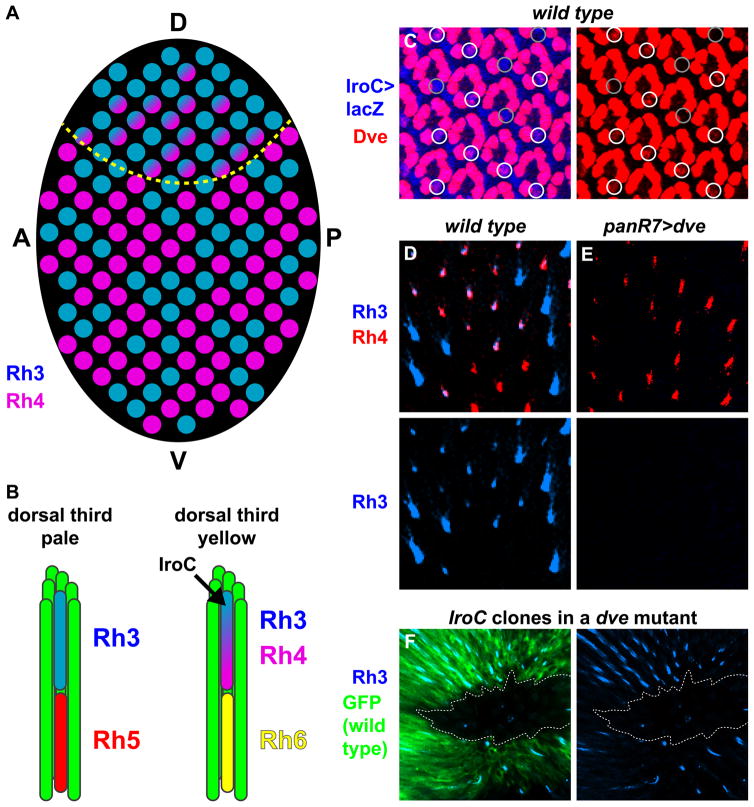
How complex networks of activators and repressors lead to exquisitely specific cell-type determination during development is poorly understood. In the Drosophila eye, expression patterns of Rhodopsins define at least eight functionally distinct though related subtypes of photoreceptors. Here, we describe a role for the transcription factor gene defective proventriculus (dve) as a critical node in the network regulating Rhodopsin expression. dve is a shared component of two opposing, interlocked feedforward loops (FFLs). Orthodenticle and Dve interact in an incoherent FFL to repress Rhodopsin expression throughout the eye. In R7 and R8 photoreceptors, a coherent FFL relieves repression by Dve while activating Rhodopsin expression. Therefore, this network uses repression to restrict and combinatorial activation to induce cell-type-specific expression. Furthermore, Dve levels are finely tuned to yield cell-type- and region-specific repression or activation outcomes. This interlocked FFL motif may be a general mechanism to control terminal cell-fate specification.


Figures

Figure 1
Figure 1. The Dve K50-homeodomain transcription factor is expressed in yR7s and outer PRs
For A–D, phalloidin in green stains actin and marks rhabdomeres. Rhodopsin proteins are localized to the actin-rich rhabdomere structures. A. 3D projection of the fly eye containing ~800 ommatidia. B. A single ommatidium. Upper dashed box indicates a section containing R7 and outer PRs. Lower dashed box indicates a section containing R8 and outer PRs. C. A section containing R7 and outer PR (R1–6) rhabdomeres. D. A section containing R8 and outer PR (R1–6) rhabdomeres. For E–G, green indicates rhabdomeres; gray indicates nuclei and cell bodies. For F–G, schematic adapted from (Wolff and Ready, 1993). E. Schematic of 1B. F. Schematic of section containing R7 and outer PRs (as in 1C). G. Schematic of section containing R8 and outer PRs (as in 1D). For H–I, left: regulatory network controlling Rh expression; middle: schematic of Rh expression in the rhabdomeres of R7 and R8; right: schematic of Rh expression in sections (top=R7 layer, bottom=R8 layer as in 1F–G). H. Pale ommatidial subtype. Rh3 expression in pR7s is coupled with Rh5 expression in pR8s. In the absence of Ss, Rh3 is expressed and p fate is signaled onto R8. The signal activates Melt expression. Melted represses Wts expression. In the absence of Wts, Rh6 is repressed and Rh5 is expressed. I. Yellow ommatidial subtype. Rh4 expression in yR7s is coupled with Rh6 expression in yR8s. Ss activates Rh4 expression and represses Rh3 and the signal. In absence of the signal, Melt is not repressed allowing for Wts expression. Wts activates Rh6 and represses Rh5. For J–K, Phalloidin (Phal) in green stains actin and marks all PR rhabdomeres. J. Rh3 and Rh4 are expressed in exclusive subsets of R7s in adults. K. Rh5 and Rh6 are expressed in exclusive subsets of R8s in adults. For L–M, left, Dve expression in adults; right, schematic of Dve expression in nuclei of PRs. Dashed circle indicates R7 cell body including nucleus and rhabdomere. Image data used to identify R7 nuclei has been removed. L. Dve is not expressed in pR7s (as indicated by Rh3 in Blue in the R7 rhabdomere at the center of the ommatidium). Dve is expressed strongly in all outer PRs. M. Dve is expressed at low levels in yR7s (as indicated by Rh4 in purple in the R7 rhabdomere at the center of the ommatidium). Dve is expressed strongly in all outer PRs. See Figure S1.
Figure 2
Figure 2. Dve represses Rh3 expression in yR7s and Rh3, Rh5, and Rh6 in outer PRs
For A–D, top, Rh3 and Rh4 stains; bottom, Rh3 alone. A. Rh3 is expressed in ~35% of R7s and Rh4 is expressed in ~65% of R7s in adult wild type animals. B. dve186 null mutants display de-repression of Rh3 in all yR7s. Rh4 is still expressed in yR7s. Rh3 protein levels are lower in yR7s (co-expressing Rh4) than in pR7s, likely due to space filling in the rhabdomere. In dve mutants, transcription of rh3 occurs at similar rates in pR7s and yR7s as assessed with a Rh3prom>GFP reporter transgene (data not shown). C. Dve is sufficient to repress Rh3 in pR7s. D. Quantification of wild type and dve186 mutant Rh3 and Rh4 expression in R7s. For top panel, error bars indicate standard deviation for the ratio of the subtypes of R7s. For bottom panel, error bars indicate standard deviation for the frequency of Rh3 expression in R7s overall. For E–G, dve null mutants display de-repression of Rh3, Rh5, and Rh6 in random subsets of outer PRs at 0 (left), 2 (middle), and 4 (right) weeks. E. Rh3 F. Rh5 G. Rh6 H. All combinations of Rh expression are observed in outer PRs in dve mutants. Top, Rh3 expression in R1. 2nd panel, Rh5 expression in R1, R5, R6. 3rd panel, Rh6 expression in R1, R3, R4, R6. Bottom, overlay, expression of Rh3, Rh5, and Rh6. I. Cell-specific expression profiles of dve mutants at 2 weeks. J, K, L indicate one example ommatidium. Left, image. Right, image with cell identification overlay. J. Phalloidin stains actin and marks all PR rhabdomeres. Phallodin staining was used to perform cell identification. K. Rh5 expression. L. Contour of Rh5 expression used to evaluate frequency of expression. M. Temporal dynamics of Rh expression. N. Dve is sufficient to repress Rh5 and Rh6 in R8s at 6 weeks (Dve gain-of-function GFP+ clone in blue). Left, triple stain; Right, Rh5 and Rh6 alone. See Figure S2.
Figure 3
Figure 3. Otd and Dve interact in an incoherent feedforward loop to control Rh expression
For A–H, images are from 2 week old adult retinas. A. Rh3 and Rh4 are expressed in exclusive subsets of R7s in wild type animals. B. Rh3 is de-repressed in yR7s and outer PRs in dve mutants. Rh4 is unaffected. C. Rh3 is not expressed in otd mutants. Rh4 is unaffected. D. Rh3 is not expressed in otd; dve double mutants. Rh4 is unaffected. E. Rh5 and Rh6 are expressed in exclusive subsets of R8s in wild type animals. F. Rh5 and Rh6 are de-repressed in outer PRs in dve mutants. G. Rh5 is not expressed in otd mutants. Rh6 is de-repressed in outer PRs in otd mutants. H. Rh5 is not expressed in otd; dve double mutants. Rh6 is de-repressed in outer PRs in otd; dve double mutants. For I–L, binding of Dve and Otd is dependent on K50 binding sites. WT=wild type promoter; M=mutation of K50 site. Arrows indicate the bands shifted upon Otd or Dve binding. Note, additional bands are observed when using the Dve protein, likely due to the presence of multiple K50 binding sites and/or multiple homeodomains within Dve yielding higher order DNA/protein structures (also see Figure S3D–F). Each of these Dve/DNA complexes is dependent upon functional K50 binding sites. I. rh3 promoter J. Dve competes with Otd for binding to the rh3 promoter. K. rh5 promoter L. rh6 promoter M. Dve expression is lost in otd null mutant tissue at 50% pupation. Left, triple stain; Right, Dve and Elav. Elav marks neuronal nuclei. N. Incoherent feedforward loop (iFFL). Left, a generalized iFFL. Right, the Od/Dve/Rh3 iFFL. For O–T, the y-axis is in arbitrary units and has been normalized to the control (promoter in the absence of Otd, Dve, and Pph13). For O–R, in all cases, Otd activates expression. Dve alone has no effect. Dve suppresses Otd-mediated activation. O. rh3 promoter P. rh5 promoter Q. rh6 promoter R. 6xK50 promoter S. rh6 promoter. Pph13 activates expression. Dve alone has no effect. Dve suppresses Pph13-mediated activation. T. rh6 promoter with mutated K50 sites. Pph13 activates expression. Dve alone has no effect. When K50 sites are mutated, Dve can not repress Pph13-mediated activation. See Figure S3.
Figure 4
Figure 4. Sal and Dve interact in a coherent feedforward loop to control Rh3 expression
A. Sal is required to repress Dve in inner PRs at 50% pupation. B. Sal is sufficient to repress Dve in outer PRs at 50% pupation. For C–K, expression assessed at 0 weeks. For C–E, dashed line indicates the equator. C. Rh3 is never expressed in outer PRs in wild type animals. D. Rh3 is expressed in a random subset of outer PRs in dve mutants. De-repression occurs more frequently in the dorsal half. E. Rh3 is expressed in nearly all outer PRs when Sal is ectopically induced in dve mutants. F. Coherent feedforward loop (cFFL). Left, a generalized cFFL. Right, the Sal/Dve/Rh3 cFFL. G. Quantification of the frequency of Rh3 expression in outer PRs from C–E. For H–J, representative examples of Rh3 expression states in R7. Thick white circles indicate R7. H. Rh3 is never expressed in R7s in sal mutants. I. Rh3 is expressed in a random subset of R7s in sal, dve double mutants. J. Rh3 is expressed in all R7s in dve mutants. K. Quantification of the frequency of Rh3 expression in R7s from H–J. For L–N, states of the interlocked feedforward loop motif. L. In the absence of Sal, Otd activates Dve which represses Rh3. M. In the absence of Sal and Dve, Otd activates Rh3 in a random subset of cells. N. In the absence of Dve and presence of Sal and Otd, Sal and Otd induce Rh3 expression in all cells. See Figure S4.
Figure 5
Figure 5. Ss activates Dve to repress Rh3 in yR7s
For A–E, white circles indicate R7s expressing Dve, gray circles indicate R7s lacking Dve. A. Dve and Ss are expressed in yR7s at 50% pupation. B. Dve is expressed in yR7s at 50% pupation. C. Dve expression is lost in R7s in ss null mutants at 50% pupation. D. Driving Ss expression in all R7s induces Dve expression at 83% pupation. E. Dve expression is lost in R7 cells mutant for ss. GFP (Green) indicates ss mutant cells. F. Rh3 is expressed in all R7s in ss mutants. G. Dve is sufficient to repress Rh3 in ss mutants. See Figure S5.
Figure 6
Figure 6. Thresholded Dve levels control IroC-mediated activation of Rh3 expression
A. Schematic of the distribution of R7s (circles) expressing Rh3, Rh4 or Rh3/Rh4 in the retina. In the dorsal third (above the yellow dashed line), Rh3 is expressed in pR7s, whereas Rh4 and Rh3 are expressed in yR7s. In the main part of the retina (below the yellow dashed line), Rh3 and Rh4 are expressed in exclusive subsets of R7s. D=dorsal, V=ventral, A=anterior, P=posterior. B. Schematics of pale and yellow ommatidia in the dorsal third. pR7s express Rh3, whereas yR7s express Rh4 and Rh3 (induced by IroC). C. Dve is expressed in a subset of R7 cells in the dorsal third that expresses IroC>lacZ at 50% pupation. White circles indicate R7s expressing Dve, gray circles indicate R7s lacking Dve. Left, IroC>LacZ and Dve; right, Dve alone. D. Wild type expression of Rh3 in dorsal yR7s expressing Rh4. E. High levels of Dve are sufficient to repress Rh3 in yR7s in the dorsal third. F. IroC mutant clones display a strong but incomplete decrease in the frequency of rh3 expression in a dve mutant background. GFP in green marks wild type tissue; non-GFP indicates IroC mutant tissue. Left, Rh3 and GFP; right, Rh3 alone.
Figure 7
Figure 7. Models
Logic of Rh regulation. Blue indicates regulatory interaction added in each level of specification. Below, we outline the specific regulatory logic for each Rh. Rh3: (A) Otd activates random Rh3 expression. (B) Otd also activates Dve which represses Rh3 in all PRs. (C) Sal represses Dve and acts together with Otd to induce Rh3 expression in all inner PRs. (F) Sens represses Rh3 in R8s, limiting expression to R7s (E). (H) Dve represses Rh3 in yR7s, restricting Rh3 expression to pR7s (G). (K) IroC in the dorsal third of the eye acts with Otd and Sal to induce Rh3 expression in yR7s despite the presence of low levels of Dve. Rh5: Similar to Rh3, (A) Otd activates random Rh5 expression. (B) Otd also activates Dve which represses Rh5 in all PRs. (C) Sal represses Dve in inner PRs allowing for de-repression of Rh5. (C) An unknown inner PR-specific factor, together with Otd, activates Rh5 in all inner PRs. (E) Pros represses Rh5 in R7s, limiting expression to R8s (F). (J) The Wts pathway represses Rh5 in yR8s leading to exclusive expression in pR8s (I). Rh6: Unlike Rh3 and Rh5, (A) random Rh6 expression is not activated by Otd, but rather requires Pph13 (likely redundantly with Otd; indicated with a dotted line). (B) Otd activates Dve which represses Rh6 in all PRs. (C) Sal represses Dve in inner PRs allowing for de-repression of Rh6. (E) Pros represses Rh6 in R7s. (F) Sens activates expression of Rh6 in all R8s. (I) Inactivation of the Wts pathway excludes Rh6 from pR8s, limiting Rh6 to yR8s (J). See Table S1.

References

    1. Alon U. Network motifs: theory and experimental approaches. Nat Rev Genet. 2007;8:450–461. - PubMed
    1. Briscoe AD. Reconstructing the ancestral butterfly eye: focus on the opsins. J Exp Biol. 2008;211:1805–1813. - PubMed
    1. Carr M, Hurley I, Fowler K, Pomiankowski A, Smith HK. Expression of defective proventriculus during head capsule development is conserved in Drosophila and stalk-eyed flies (Diopsidae) Dev Genes Evol. 2005;215:402–409. - PubMed
    1. Chou WH, Huber A, Bentrop J, Schulz S, Schwab K, Chadwell LV, Paulsen R, Britt SG. Patterning of the R7 and R8 photoreceptor cells of Drosophila: evidence for induced and default cell-fate specification. Development. 1999;126:607–616. - PubMed
    1. Cook T, Pichaud F, Sonneville R, Papatsenko D, Desplan C. Distinction between color photoreceptor cell fates is controlled by Prospero in Drosophila. Dev Cell. 2003;4:853–864. - PubMed

Publication types

MeSH terms

LinkOut - more resources