. 2011 Jun 10;145(6):969-80.
doi: 10.1016/j.cell.2011.05.022.

Riboneogenesis in yeast

Affiliations

Riboneogenesis in yeast

Michelle F Clasquin et al. Cell. .

Abstract

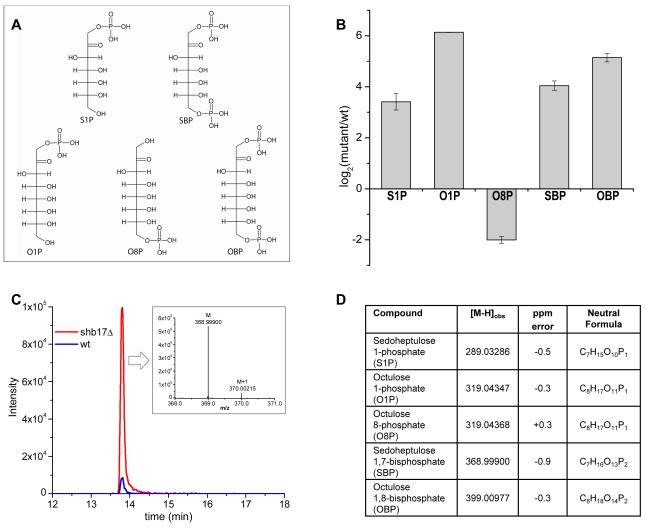
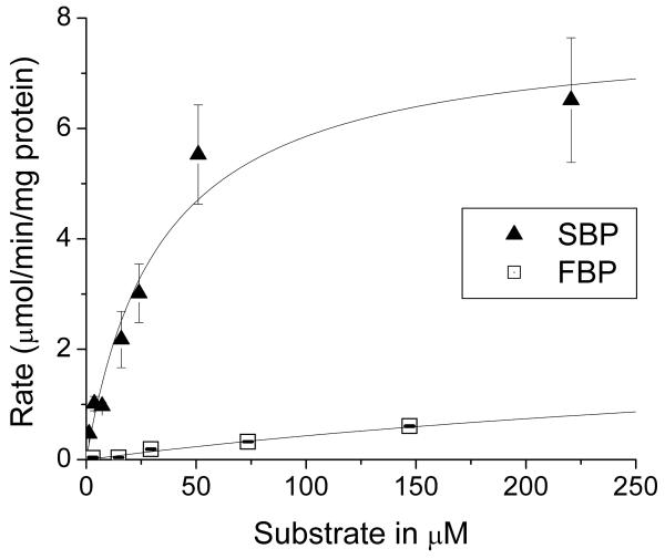
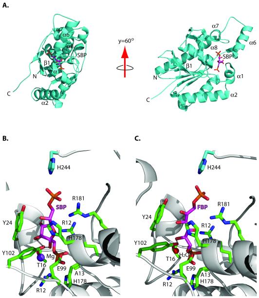
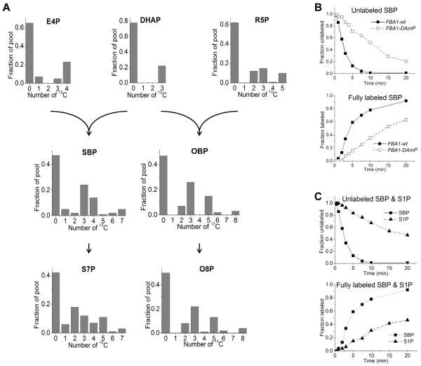
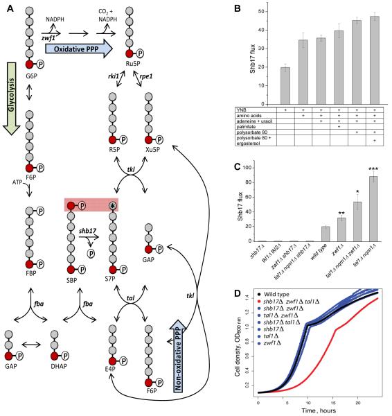
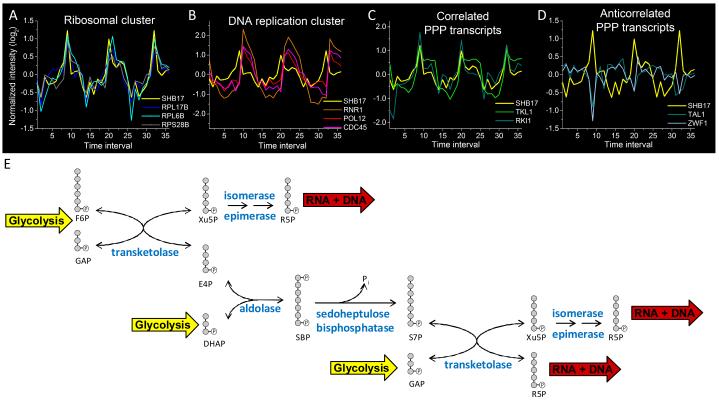
Glucose is catabolized in yeast via two fundamental routes, glycolysis and the oxidative pentose phosphate pathway, which produces NADPH and the essential nucleotide component ribose-5-phosphate. Here, we describe riboneogenesis, a thermodynamically driven pathway that converts glycolytic intermediates into ribose-5-phosphate without production of NADPH. Riboneogenesis begins with synthesis, by the combined action of transketolase and aldolase, of the seven-carbon bisphosphorylated sugar sedoheptulose-1,7-bisphosphate. In the pathway's committed step, sedoheptulose bisphosphate is hydrolyzed to sedoheptulose-7-phosphate by the enzyme sedoheptulose-1,7-bisphosphatase (SHB17), whose activity we identified based on metabolomic analysis of the corresponding knockout strain. The crystal structure of Shb17 in complex with sedoheptulose-1,7-bisphosphate reveals that the substrate binds in the closed furan form in the active site. Sedoheptulose-7-phosphate is ultimately converted by known enzymes of the nonoxidative pentose phosphate pathway to ribose-5-phosphate. Flux through SHB17 increases when ribose demand is high relative to demand for NADPH, including during ribosome biogenesis in metabolically synchronized yeast cells.


Figures

Figure 1
Figure 1
Metabolomic phenotype of shb17Δ. (A) Metabolite structures associated with metabolic phenotype of shb17Δ. For fragmentation data confirming compound structures, see Supplemental Figure 1. (B) Relative quantitation of metabolites. Data shown is arithmetic mean ± SE of N=4 independent biological replicates. (C)The negative ionization mode extracted ion chromatogram for SBP in shb17Δ and wild type S. cerevisiae. Inset: Mass spectrum displaying the accurate mass for the parent ion (M) and natural 13C abundance ion (M+1) observed for SBP in negative ionization mode via LC/Exactive Orbitrap MS. (D) Table of [M-H] ions with altered abundance between shb17Δ and wild type.
Figure 2
Figure 2
Hydrolysis of SBP and FBP by purified recombinant Shb17: dependence on substrate concentration. Data are the mean ± standard error for two independent experiments. Full data for FBP are available in Supplemental Figure 2.
Figure 3
Figure 3
Structure of the Shb17/SBP complex. (A) Overall fold of the Shb17 (H13A) in complex with SBP (PDB 3OI7, grey ribbon) shown in two orientations with secondary structural elements being labeled. The SBP molecule (magenta carbon atoms) is shown in a stick representation. (B) Close-up view of the active site of Shb17 in complex with SBP. The side chains of residues in contact with SBP are displayed in a stick representation (green carbon atoms) and labeled. SBP is shown in a stick representation (magenta carbon atoms) and labeled, whereas the Mg2+ ion is shown as a purple sphere and labeled. (C) Active site of Shb17 in complex with FBP, a similar view as (B). The red sphere denotes a water molecule. Y102 makes two hydrogen bonds with SBP, whereas only one hydrogen bond can be formed between this residue and FBP. These hydrogen bonds are shown by dashed lines in parts B and C.
Figure 4
Figure 4
SBP and OBP are synthesized in vivo by C3 + C4 and C3 + C5 subunits via fructose bisphosphate aldolase. (A) Cells were switched from unlabeled to 70:30 unlabeled glucose:[U-13C]-glucose. Labeling patterns of erythrose-4-phosphate (E4P), dihydroxyacetone-phosphate (DHAP), ribose-5-phosphate (R5P), SBP and OBP were measured in shb17Δ, where SBP and OBP accumulate and hence are more readily quantitated. The reaction products sedoheptulose-7-phosphate (S7P) and octulose 8-phosphate (O8P) were measured in wild type (for data on S7P in shb17Δ see Supplementary Figure 4A). Labeling is reported 20 minutes after nutrient switch for all compounds except OBP, where data is taken at 120 min due to its slower labeling. (B) Kinetics of labeling of SBP after switching shb17Δ cells with wild type fructose bisphosphate aldolase (FBA1-wt), or the Decreased Abundance by mRNA Perturbation allele (FBA1-DAmP) into [U-13C6]-glucose. For associated pool size and kinetic data, see Supplemental Figure 4 (B-C). (C) Kinetics of labeling of SBP and S1P after switching shb17Δ cells into [U-13 C6]-glucose.
Figure 5
Figure 5
Shb17 feeds carbon into the non-oxidative pentose phosphate pathway. (A) Flux through Shb17 into S7P can be measured using [6-13C1]-glucose. [6-13C1]-glucose leads to [7-13C1]-S7P when S7P is made via the oxidative PPP or the non-oxidative PPP. However, when S7P is produced from SBP via Shb17, a fraction of the S7P pool is doubly labeled: [1,7-13C2]-S7P. Flux is calculated based on the measured isotopic distribution of SBP and S7P. (B) Flux through Shb17 is increased by supplementation with nutrients whose endogenous production requires NADPH, and thus drives oxidative PPP flux. All measurements are performed in wild type yeast. YNB is yeast nitrogen base without amino acids plus 2% glucose. Supplementation with amino acids includes 17 amino acids. Data shown is the arithmetic mean ± SE of N=3 technical replicates. (C) Effects of PPP gene deletions on Shb17 flux. Deletions are: glucose 6-phosphate dehydrogenase zwf1Δ; transketolase tkl1Δ/ tkl2Δ; transaldolase is tal1Δ/nqm1Δ. Less than 1% doubly labeled S7P was observed in any shb17Δ strain in all measured conditions. All strains were grown in YNB + 2% glucose and supplements as required: methionine for zwf1Δ; synthetic complete media including aromatic amino acids for tkl1Δ/tkl2Δ. (C) Triple deletion of the sedoheptulose bisphosphatase SHB17, the glucose-6-phosphate dehydrogenase ZWF1, and the transaldolase TAL1, causes a growth defect. Optical density was measured during growth at 30 degrees C in YPD. Growth data are presented in Supplemental Table 3.
Figure 6
Figure 6
SHB17 expression cycles in concert with the yeast metabolic cycle. (A)-(D) A time series of gene expression during the ~300 minute yeast metabolic cycle is plotted from data presented in (Tu et al., 2005), where each time interval represents ~25 minutes. (A) SHB17 is co-expressed with ribosomal transcripts (shown here: two components of the 60S ribosomal subunit, RPL17B and RPL6B, and one of the 40S subunit, RPS28B). (B) Ribosomal protein transcript expression precedes transcripts associated with DNA replication (shown here: ribonucleotide reductase, RNR1, the B subunit of DNA polymerase, POL12, and a DNA replication initiation factor, CDC45). (C) SHB17 expression correlates with selected PPP transcripts including transketolase (TKL1) and ribose 5-phosphate ketol-isomerase (RKI1). (D) SHB17 expression is anticorrelated with other PPP transcripts including transaldolase (TAL1) and glucose 6-phosphate dehydrogenase (ZWF1). Y-axis displays log2 transformed intensity, with each gene median centered at 0. (E) Riboneogenic pathway in yeast. The expression data in (A)-(D) suggest a coordinated role of the transketolase TKL1, the ribose ketoisomerase RKI1, and the sedoheptulose bisphosphatase SHB17 in riboneogenesis. The aldolase FBA1 is constituively expressed, consistent with its central role in both glycolysis and gluconeogenesis. The ribulose epimerase RPE1 is also continually expressed. Together, the enzymes work to shunt glycolytic intermediates to ribose. The overall scheme converts one hexose-P and three triose-P to three pentose-P units.

References

    1. SGD project [November 16, 2010];Saccharomyces Genome Database. http://www.yeastgenome.org/
    1. Allen J, Davey HM, Broadhurst D, Heald JK, Rowland JJ, Oliver SG, Kell DB. High-throughput classification of yeast mutants for functional genomics using metabolic footprinting. Nat Biotechnol. 2003;21:692–696. - PubMed
    1. Amberg DC, Burke DJ, Strathern JN. Methods in Yeast Genetics. Cold Spring Harbor Laboratory Press; Cold Spring Harbor, NY: 2005.
    1. Bartlett GR, Bucolo G. Octulose phosphates from the human red blood cell. Biochem Biophys Res Commun. 1960;3:474–478. - PubMed
    1. Bartlett GR, Bucolo G. The metabolism of ribonucleoside by the human erythrocyte. Biochim Biophys Acta. 1968;156:240–253. - PubMed

Publication types

MeSH terms

Substances

Associated data