Review
. 2011 Dec 5;347(1-2):80-9.
doi: 10.1016/j.mce.2011.05.017. Epub 2011 Jun 1.

Vitamin D metabolism and function in the skin

Affiliations
Review

Vitamin D metabolism and function in the skin

Daniel D Bikle. Mol Cell Endocrinol. .

Abstract

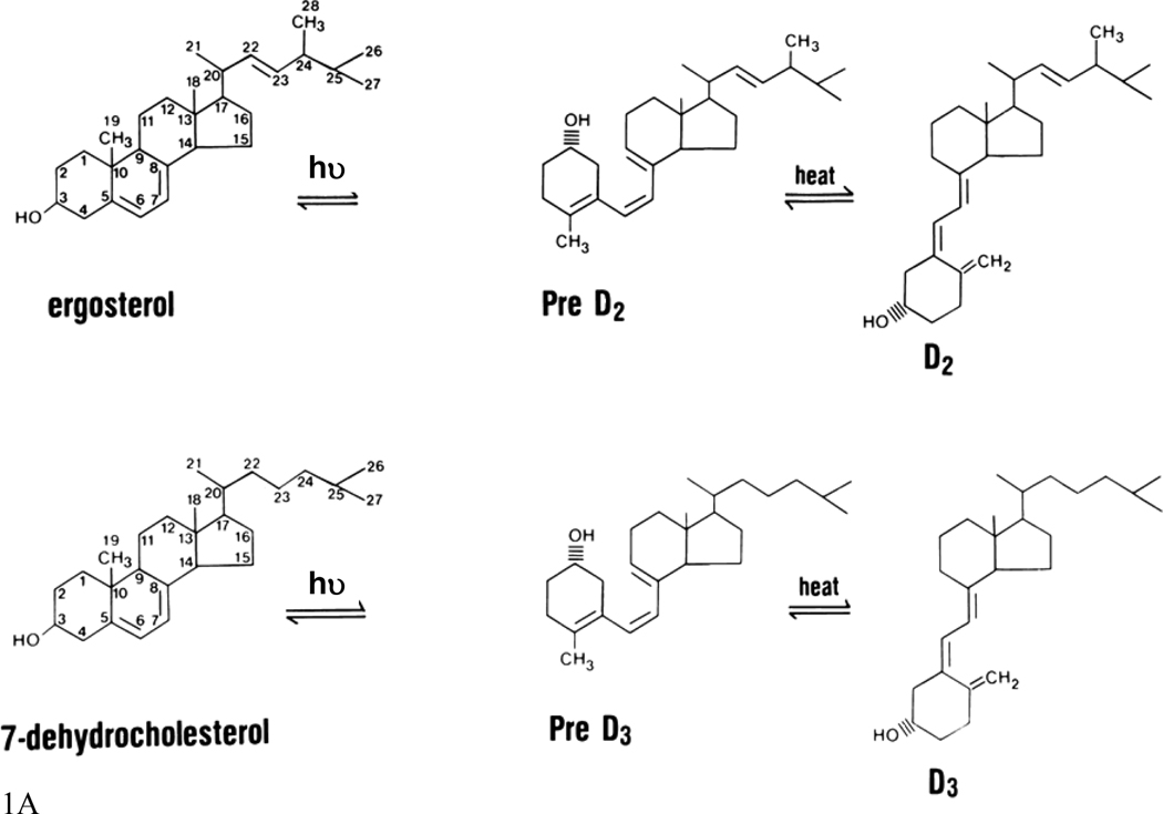
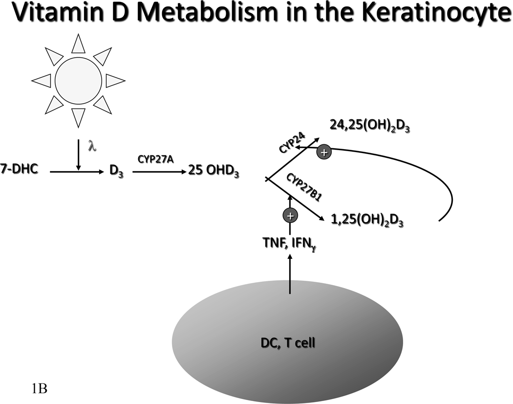
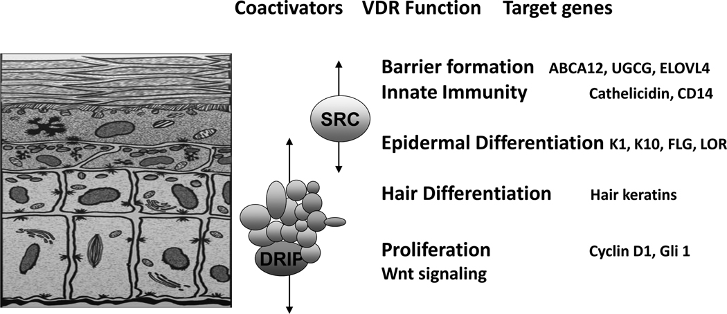
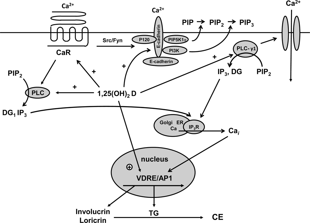
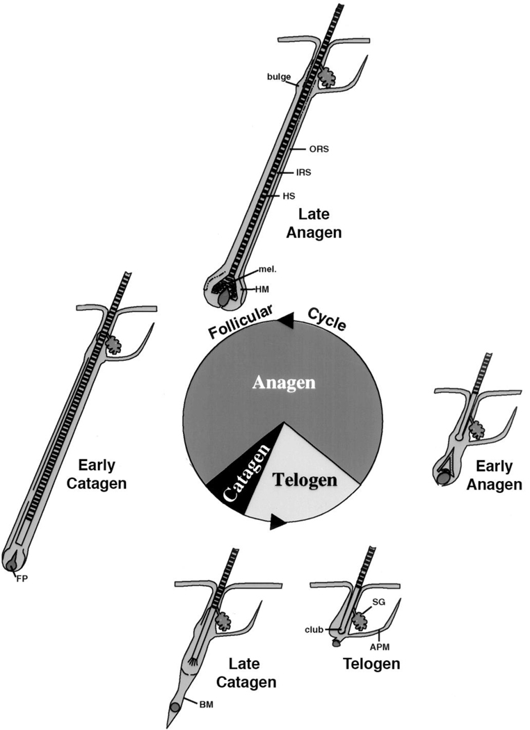
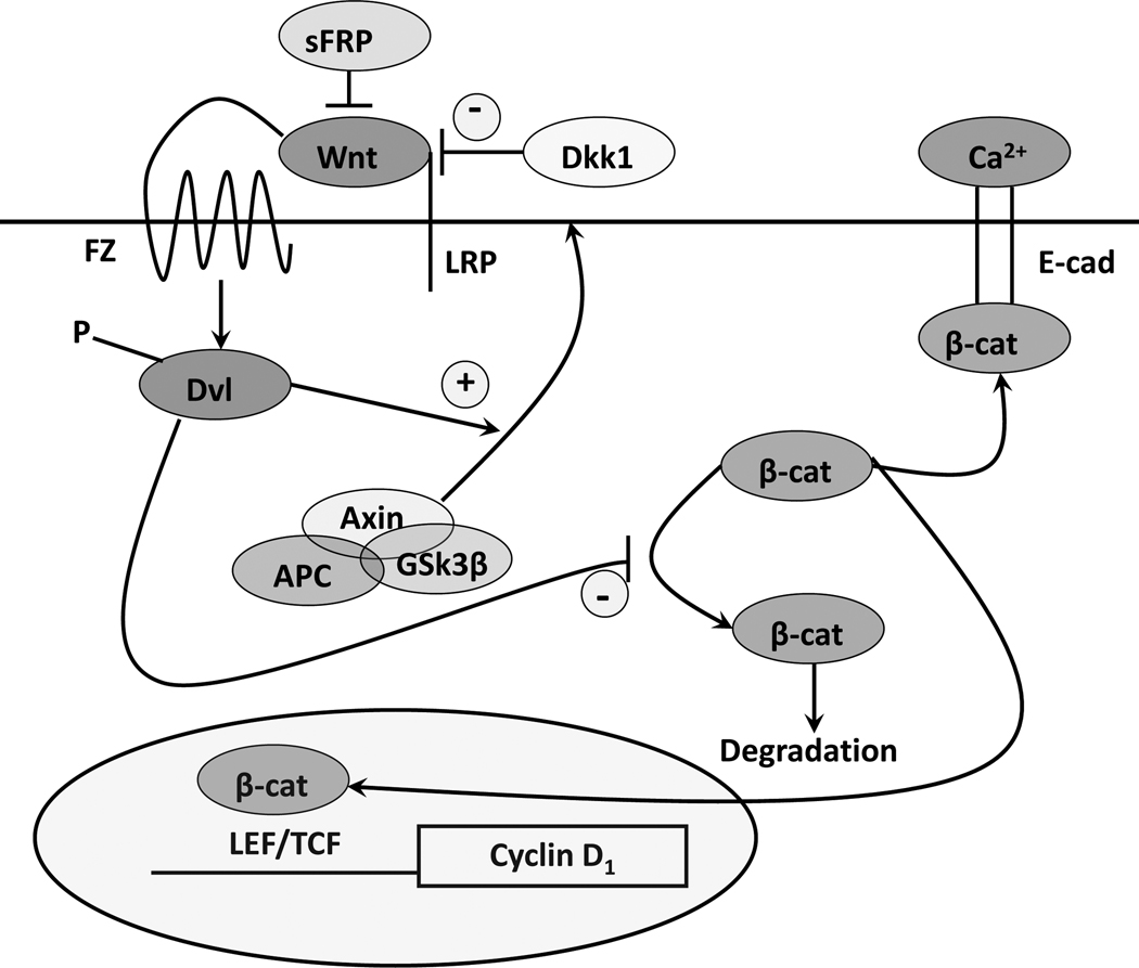
The keratinocytes of the skin are unique in being not only the primary source of vitamin D for the body, but in possessing the enzymatic machinery to metabolize vitamin D to its active metabolite 1,25(OH)(2)D. Furthermore, these cells also express the vitamin D receptor (VDR) that enables them to respond to the 1,25(OH)(2)D they produce. Numerous functions of the skin are regulated by 1,25(OH)(2)D and/or its receptor. These include inhibition of proliferation, stimulation of differentiation including formation of the permeability barrier, promotion of innate immunity, and promotion of the hair follicle cycle. Regulation of these actions is exerted by a number of different coregulators including the coactivators DRIP and SRC, the cosuppressor hairless (Hr), and β-catenin. This review will examine the regulation of vitamin D production and metabolism in the skin, and explore the various functions regulated by 1,25(OH)(2)D and its receptor.


Figures

Figure 1
Figure 1. The production of vitamin D and its subsequent metabolism
A. Vitamin D production. Sunlight (the ultraviolet B component) breaks the B ring of the precursor sterol to form pre- D2 or pre-D3 from ergosterol or 7-dehydrocholesterol, respectively. In a temperature dependent step the A ring is rotated around the C5 to C6 double bond so that the 3β-hydroxyl group is positioned below the plane of the A ring to form D2 and D3. B. Metabolism of Vitamin D in the keratinoocyte. In the rest of the body the liver converts vitamin D to 25OHD. The kidney converts 25OHD to 25OHD and 1,25(OH)2D. Regulation of CYP27B1 in the kidney is exerted by calcium, phosphorus, parathyroid hormone, FGF23, and 1,25(OH)2D itself. However, in the keratinocyte, vitamin D can be directly metabolized to 25OHD and further metabolized to 25OHD and 1,25(OH)2D. Cytokines such as TNF and IFN-γ are the principal regulators of CYP27B1 in the keratinocyte.
Figure 2
Figure 2. The different layers of the epidermis, and the functions within those layers regulated by VDR and its coactivators
The basal layer of the epidermis (stratum basale) contains the stem cells that through proliferation provide the cells for the upper layers. As the keratinocytes leave the basal layer differentiation takes place with K1, K10, involucrin, and transglutaminase being expressed in the stratum spinosum, filaggrin and loricrin being expressed in the stratum granulosum. Lamellar bodies forming in the stratum granulosum inject their lipid content into the intercellular spaces between the stratum granulosum and stratum corneum to provide the water proofing for the permeability barrier. DRIP205 is most highly expressed in the stratum basale and spinosum where it participates with VDR in regulating proliferation in partnership with the wnt/β-catenin signaling pathway. Cyclin D1 and Gli 1 (a transcriptional factor for the hedgehog pathway) are regulated by VDR, DRIP205, and β-catenin. SRC3 on the other hand is found in highest concentration in the stratum granulosum where it participates with VDR in the regulation of terminal differentiation. SRC3 is critical for lipid processing and lamellar body formation required for formation of the permeability barrier as well as 1,25(OH)2D induction of cathelicidin and CD14 required for innate immunity.
Figure 3
Figure 3. Calcium and 1,25(OH)2D interactions in the regulation of keratinocyte differentiation
Critical steps in calcium induced keratinocyte differentiation involve activation of the calcium sensing receptor (CaR) and formation of the E-cadherin complex at the membrane. This complex includes a number of catenins and phospholipid modifying enzymes (PI3K, PIP5K1α) that are critical for subsequent differentiation events. In particular the activation of phospholipase C-γ1 (PLC-γ1), the enzyme largely responsible for maintaining increased levels of intracellular calcium (Cai) through its effects on both plasma membrane calcium channels and in the production of IP3 from PIP2 for stimulation of calcium release from intracellular stores, requires the formation of the E-cadherin complex. Hydrolysis of PIP2 by PLC-γ1 also leads to diacylglycerol production (DG) and activation of protein kinases C, that also play an important role in keratinocyte differentiation. Within hours of the calcium switch keratinocytes change from making the basal keratins K5 and K14 to making keratins K1 and K10 followed, subsequently, by increased levels of profilaggrin, involucrin and loricrin. Loricrin, involucrin and other proteins are cross linked into the insoluble cornified envelope by the calcium sensitive, membrane bound form of transglutaminase, which like involucrin and loricrin increases within 24 hours after the calcium switch. The induction of these proteins represents a genomic action (likely indirect) of calcium as indicated by a calcium induced increase in mRNA levels and transcription rates. The CaR by regulating phospholipase C (PLC) activity controls the production of inositol tris phosphate (IP3) and diacyl glycerol (DG). PLC-β is activated directly by CaR via a G protein coupled mechanism, whereas PLC-γ1 is activated by phosphatidyl inositol tris phosphate (PIP3), levels that are maintained in the membrane by phosphatidyl inositol 3 kinase (PI3K) bound to the E-cadherin complex. CaR regulates E-cadherin complex formation through src/fyn tyrosine kinases that phosphorylate the catenins and PI3K essential for their binding to E-cadherin. Extracellular calcium (Cao) activates these processes through the CaR and by stabilizing E-cadherin membrane localization. 1,25(OH)2D modulates calcium regulated differentiation at several steps. First, 1,25(OH)2D increases CaR expression, thus making the cell more responsive to calcium. Second, 1,25(OH)2D induces all the PLCs, as does calcium, again increasing the responsiveness of the cell to calcium. Third, the VDR is required for calcium induced formation of the E-cadherin complex in the membrane, in part through induction of E-cadherin as well as CaR. Finally, 1,25(OH)2D induces the transcription of genes such as involucrin and transglutaminase and possibly the other differentiation markers in addition to PLC and CaR.
Figure 4
Figure 4. Hair follicle cycling
The initial developmental phase of hair follicle development terminates with catagen and the first telogen, after which repetitive cycles of anagen (the growth phase), catagen (the regression phase), and telogen (the resting phase) occur throughout the life span of the animal. In general the hair follicle spends most of its time in anagen. But cycle duration varies according to location, gender, age, species. The bulge is the source of stem cells for the regenerating hair follicle, responding to signals from the dermal or follicular papilla (FP). Although VDR is not required for the developmental phase of the hair follicle, it is essential for subsequent hair follicle cycling. ORS: outer root sheath, IRS: inner root sheath, HS: hair shaft, mel: melanin for the hair shaft, HM: hair matrix, BM: basement membrane, SG: sebaceous gland, APM: arrector pili muscle. Figure adapted from figure 1 in KS Stenn and R Paus, Physiol Rev:81:449–494, 2001.
Figure 5
Figure 5. The canonical wnt signaling pathway
Wnts bind to their frizzled receptors (FZ) and coreceptors LRP in the membrane. This binding can be blocked by dickkopf (Dkk) or soluble frizzled related proteins (sFRP). Activation of FZ by wnt results in phosphorylation of disheveled (Dvl), which induces the disruption of the axin/APC/GSK-3β complex and recruitment of axin to the membrane. This complex when active phosphorylates β-catenin, leading to its proteosomal degradation. However, following wnt stimulation β-catenin is no longer degraded and can enter the nucleus where in combination with members of the LEF/TCF family can induce expression of its target genes such as cyclin D1. β-catenin also binds to the E-cadherin complex in the plasma membrane, binding which promotes differentiation. 1,25(OH)2D/VDR interact with wnt signaling at multiple points. 1,25(OH)2D/VDR binds β-catenin reducing its transcriptional activity. 1,25(OH)2D/VDR promotes the formation of the E-cadherin/catenin complex in the membrane also serving to reduce translocation of β-catenin into the nucleus.

References

    1. Acevedo ML, Lee KC, Stender JD, et al. Selective recognition of distinct classes of coactivators by a ligand-inducible activation domain. Mol Cell. 2004;13:725–738. - PubMed
    1. Ahmad W, Faiyaz ul Haque M, Brancolini V, et al. Alopecia universalis associated with a mutation in the human hairless gene. Science. 1998;279:720–724. - PubMed
    1. Bell NH, Greene A, Epstein S, et al. Evidence for alteration of the vitamin D-endocrine system in blacks. Journal of Clinical Investigation. 1985;76:470–473. - PMC - PubMed
    1. Bienz M. beta-Catenin: a pivot between cell adhesion and Wnt signalling. Curr Biol. 2005;15:R64–R67. - PubMed
    1. Bikle DD, Nemanic MK, Gee E, et al. 1,25-Dihydroxyvitamin D3 production by human keratinocytes. Kinetics and regulation. J Clin Invest. 1986a;78:557–566. - PMC - PubMed

Publication types