. 2011 Aug 15;417(1):113-25.
doi: 10.1016/j.virol.2011.05.007. Epub 2011 Jun 12.

Long-distance kissing loop interactions between a 3' proximal Y-shaped structure and apical loops of 5' hairpins enhance translation of Saguaro cactus virus

Affiliations

Long-distance kissing loop interactions between a 3' proximal Y-shaped structure and apical loops of 5' hairpins enhance translation of Saguaro cactus virus

Maitreyi Chattopadhyay et al. Virology. .

Abstract

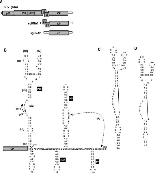
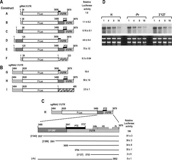
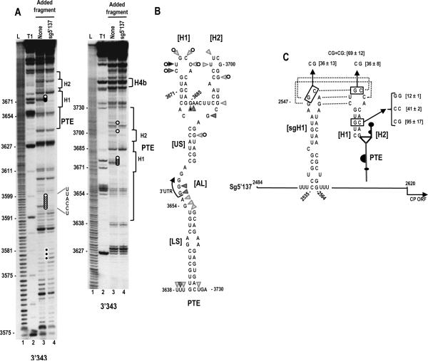
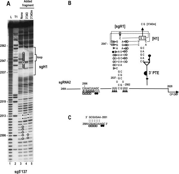
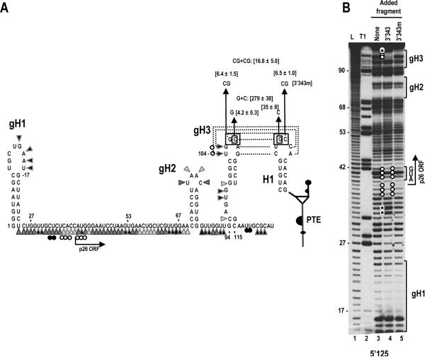
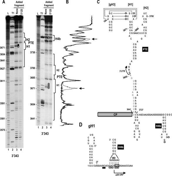
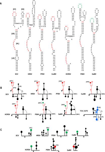
Circularization of cellular mRNAs is a key event prior to translation initiation. We report that efficient translation of Saguaro cactus virus (SCV) requires a 3' translational enhancer (PTE) located partially in coding sequences. Unlike a similar PTE reported in the 3' UTR of Pea enation mosaic virus that does not engage in an RNA:RNA interaction (Wang Z. et al., J. Biol. Chem. 284, 14189-14202, 2009), the SCV PTE participates in long distance RNA:RNA interactions with hairpins located in the p26 ORF and in the 5' UTR of one subgenomic RNA. At least two additional RNA:RNA interactions are also present, one of which involves the p26 initiation codon. Similar PTE can be found in six additional carmoviruses that can putatively form long-distance interactions with 5' hairpins located in comparable positions.


Figures

Fig. 1
Fig. 1
The SCV system. (A) Genome organization of SCV. SCV gRNA contains five ORFs. The viral replicase p86 is the ribosomal read -through product of p26. sgRNA1 is a bi-cistronic mRNA for expression of p6 and p9, which are movement proteins. sgRNA2 codes for the capsid protein (CP). (B) Putative secondary structure in the 3' UTR of SCV. The PTE is partially within the CP ORF (gray sequence). Pr, H5 and pseudoknot ψ1 are conserved in carmoviruses. Most carmoviruses also have H4b. H5 and H4b in TCV are part of the TCV TSS. US, upper stem; AL, asymmetric loop; LS, lower stem. (C) The similar PTE of PEMV, which is known to bind to translation initiation factor eIF4E (Wang et al., 2009). (D) The PTE of PMV, previously determined to be a 3'TE (Batten et al., 2006).
Fig. 2
Fig. 2
Identifying important translation sequences in SCV. (A) gRNA constructs contained either the gRNA 5' UTR (1–39 nt) or extended 5' sequence (1–80 or 1–125 nt) at the 5' end and either the exact 3' UTR (positions 3656–3879), extended 3' sequence (positions 3480–3879; 400 nt), or 223 nt of random sequence at the 3' end. Short black box denotes partial PTE element, with the larger black box labeled “PTE” denoting the full-sized PTE spanning the 3' UTR junction. Light grey regions are SCV UTR sequences and dark grey regions are SCV coding sequences. Transcripts were inoculated into A. thaliana protoplasts along with a control R-Luc construct and luciferase levels assayed 18 h later. Standard deviation in three replicate experiments is shown. (B) sgRNA2 constructs contained the exact 5' UTR (positions 2484–2620) at the 5' end and either the 3' UTR, 3' 400 nt, or 400 random nt at the 3' end. (C) Deletions were constructed in construct H, containing the 5' UTR of sgRNA2 and 3' 400 nt. Black lines represent remaining sequence. Identities of three fragments (3'343, 3'289 and -Pr) used in subsequent experiments are denoted. Positions of PTE, H4b, H5 and Pr are indicated. Standard deviation in three replicate experiments is shown. (D) Stability of parental construct H and deletion constructs –Pr and 3'127 in protoplasts. Transcribed RNAs were transfected into protoplasts and total RNA isolated at the times (in hours) indicated above each lane. RNA was subjected to RNA gel blot analysis using oligonucleotide probes complementary to the luciferase coding region.
Fig. 3
Fig. 3
A kissing loop interaction exists between the 3' PTE and sgRNA2 5' UTR hairpin sgH1 that contributes to 3' translational enhancement. (A) In-line probing of fragment 3'343 in the absence and presence of the sgRNA2 5' UTR (5'137). The panel on the right is a longer run of the samples shown in the left panel. Fragment 3'343 was labeled at its 5' end and allowed to self cleave at 25°C for 14 h. Intensity levels of the bands in lanes 3 and 4 are proportional to the flexibility of the residues. L, alkaline-generated ladder; T1, RNase T1 digest of partially denatured RNA. Note loss of flexibility in the PTE H1 loop in the presence of fragment 5'137, which is consistent with base-pairing between the PTE H1 apical loop and PTE-complementary sequence in the sgH1 apical loop (see C). (B) Flexibility of residues in the 3' PTE region. Data is from lane 3 in (A). Darker triangles denote higher flexibility. Open circles reflect residues that lose flexibility in the presence of fragment 5'137. (C) Single and compensatory mutations generated in construct H that disrupt or reform the PTE H1 stem or the PTE RNA:RNA interaction. Levels of translation as a percentage of wt construct H are shown. Results are from three experiments with standard deviations given. Sequences that can putatively pair are connected by dotted lines.
Fig. 4
Fig. 4
Effect of the PTE kissing loop interaction on the structure of the sgRNA2 5' UTR in the vicinity of sgH1. (A) In-line probing of the sgRNA2 5' UTR (5'137) in the absence and presence of fragment 3'343 and 3'343 with mutations disrupting the RNA:RNA interaction (3'343m). The 5'137 fragment was labeled at its 5' end and allowed to self cleave at 25°C for 14 h in the absence and presence of 3'343 or 3'343m. L, alkaline-generated ladder; T1, RNase T1 digest of partially denatured RNA. Open and closed circles denote residues that lose or gain flexibility, respectively, in the presence of wt 3'343 or 3'343m. Note enhanced stability of the residues in the sgH1 stem and loop when 5'137 and wt 3'343 are combined, which is consistent with base-pairing between sgH1 and PTE. Additional changes are also found in an upstream region. (B) Flexibility of residues in the sgH1 region of the sgRNA2 5' UTR. Data is from (A). Darker triangles denote higher flexibility. Open and closed circles denote residues that lose or gain flexibility, respectively, in the presence of wt 3'343. Sequences that can putatively pair are connected by dotted lines. The mutations in 3'343m that disrupt the RNA:RNA interaction are shown. (C) Possible pairing between sequence at positions 3551–3557 in the 3' region of SCV and sg5'137. Open and closed circles are as designated in (B).
Fig. 5
Fig. 5
A kissing loop interaction between the 3' PTE and gRNA p26 ORF coding sequences contributes to 3' translational enhancement. (A) Putative structure of the 5' 125 nt of SCV. The secondary structure shown is based on mFold predictions and results of in-line probing (see B). The potential pairing between hairpin gH3 and the PTE H1 is shown with dotted lines connecting the paired residues. Flexibility of residues in the 5' 125 nt region is from lane 3 in (B). Darker triangles denote higher flexibility. Open and closed circles reflect residues that lose or gain flexibility, respectively in the presence of fragment 3'343. Single and compensatory mutations generated in construct E that disrupt or reform the PTE RNA:RNA interaction are shown. Levels of translation as a percentage of wt construct E are given. Results are from three experiments with standard deviations given. (B) In-line probing of labeled fragment 5'125 in the absence and presence of fragments 3'343 and 3'343m. L, alkaline-generated ladder; T1, RNase T1 digest of partially denatured RNA. Note that two gH3 loop residues lose flexibility and two residues near the base of gH3 show enhanced flexibility in the presence of fragment 3'343 only (lane 4, top), which is consistent with base-pairing between the apical loops of PTE H1 and gH3 (see A). Also note the presence of a second region of altered flexibility in the vicinity of the p26 initiation codon, which is retained in the absence of the PTE-mediated RNA:RNA interaction (lane 5) suggesting the presence of a second RNA:RNA interaction.
Fig. 6
Fig. 6
Two long-distance interactions occur between the 5' end of the SCV genomic RNA and the 3' region. (A) In-line probing of fragment 3'343 in the absence and presence of the gRNA fragment 5'125. The panel on the right is a longer run of the samples shown in the left panel. L, alkaline-generated ladder; T1, RNase T1 digest of partially denatured RNA. Note loss of flexibility in the PTE H1 loop (lane 4, five consecutive white circles) in the presence of fragment 5'125, which is consistent with base-pairing between the PTE H1 apical loop and PTE-complementary sequence in hairpin gH3 (see C). Cleavages of two residues in the H4b apical loop were also reproducibly reduced in intensity in the presence of 5'125 but not 5'137 (see Fig. 3A). (B) Densitometry tracing of the right side autoradiograph in A. Gray tracing is from lane 3 and black tracing is from lane 4. Arrows denote positions where the cleavage pattern differs between the two lanes. (C) Location of cleavage pattern differences between lanes 3 and 4 in A. Open circles reflect residues that lose flexibility in the presence of fragment 5'125. (D) Possible pairing between the apical loop of H4b and the region of reduced flexibility in the 5'125 fragment in the presence of 3'343. The start of the p26 ORF is indicated. The initiation codon is boxed.
Fig. 7
Fig. 7
Comparison of PTE and PTE-interacting sequences in carmoviruses. (A) Sequences of PTE found in seven carmoviruses. Conserved sequences are colored alike. Note that the sequence in green contains the complement of the conserved GCCA sequence (UGGC). (B) Location of the carmovirus PTE in the 3' regions of the viral genomes. Black triangle denotes location of the stop codon for the CP ORFs. The PTE is in pink, the conserved H1 sequence CUGCCA is denoted by a red terminal loop and the conserved H1 sequence UUGGCC/ G is d enoted by a green terminal loop. Positions of the conserved sequences within the viral genome is given. Note that all viruses except GaMV have Pr, ψ1, H5 and H4b and none have ψ2. Secondary structures are based on mFold predictions and tertiary structures are based on visual observations. The structure of the 3' UTR of TCV and CCFV is provided for reference. (C) Location of PTE H1 complementary sequence near the 5' end of the gRNA. Note that all sequences are in apical loops of hairpins located either at the 5' terminus or positioned such that the loop sequence is 100 to 106 nt from the 5' end as denoted in the figure. The distance of these latter complementary sequences from the initiation codons (gray triangle) is highly variable. Secondary structures are based on mFold predictions. Sequences complementary to PTE H1 sequence CUGCCA is in red and UUGGCC/ G is in green. HRSV, Honeysuckle ringspot virus; PFBV, Pelargonium flower break virus; CarMV, Carnation mottle virus; PSNV; Pea stem necrosis virus; GaMV, Galinsoga mosaic virus.

References

    1. Batten JS, Desvoyes B, Yamamura Y, Scholthof KBG. A translational enhancer element on the 3'-proximal end of the Panicum mosaic virus genome. FEBS Let. 2006;580:2591–2597. - PubMed
    1. Bradrick SS, Walters RW, Gromeier M. The hepatitis C virus 3'-untranslated region or a poly(A) tract promote efficient translation subsequent to the initiation phase. Nucl. Acids Res. 2006;34:1293–1303. - PMC - PubMed
    1. Bushell M, Sarnow P. Hijacking the translation apparatus by RNA viruses. J. Cell Biol. 2002;158:395–399. - PMC - PubMed
    1. Chiu WW, Kinney RM, Dreher TW. Control of translation by the 5'- and 3'-terminal regions of the dengue virus genome. J. Virol. 2005;79:8303–8315. - PMC - PubMed
    1. de Quinto SL, Saiz M, de la Morena D, Sobrino F, Martinez-Salas E. IRES-driven translation is stimulated separately by the FMDV 3'-NCR and poly(A) sequences. Nucl. Acids Res. 2002;30:4398–4405. - PMC - PubMed

Publication types