. 2011 Jun 10;88(6):767-777.
doi: 10.1016/j.ajhg.2011.05.007.

Mutations in PRDM5 in brittle cornea syndrome identify a pathway regulating extracellular matrix development and maintenance

Affiliations

Mutations in PRDM5 in brittle cornea syndrome identify a pathway regulating extracellular matrix development and maintenance

Emma M M Burkitt Wright et al. Am J Hum Genet. .

Erratum in

  • Am J Hum Genet. 2011 Aug 12;89(2):346

Abstract

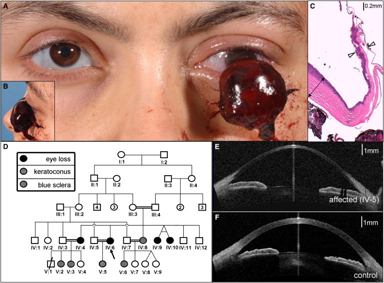
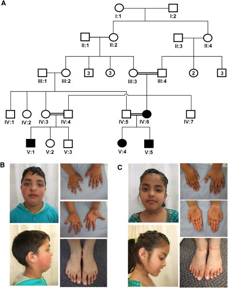
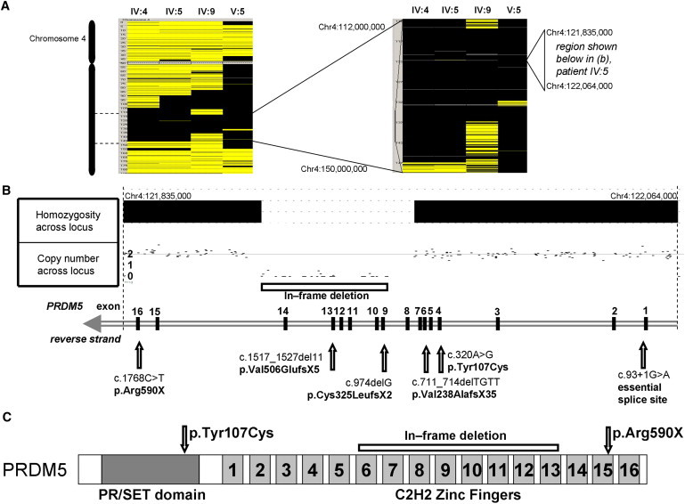
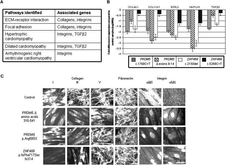
Extreme corneal fragility and thinning, which have a high risk of catastrophic spontaneous rupture, are the cardinal features of brittle cornea syndrome (BCS), an autosomal-recessive generalized connective tissue disorder. Enucleation is frequently the only management option for this condition, resulting in blindness and psychosocial distress. Even when the cornea remains grossly intact, visual function could also be impaired by a high degree of myopia and keratoconus. Deafness is another common feature and results in combined sensory deprivation. Using autozygosity mapping, we identified mutations in PRDM5 in families with BCS. We demonstrate that regulation of expression of extracellular matrix components, particularly fibrillar collagens, by PRDM5 is a key molecular mechanism that underlies corneal fragility in BCS and controls normal corneal development and maintenance. ZNF469, encoding a zinc finger protein of hitherto undefined function, has been identified as a quantitative trait locus for central corneal thickness, and mutations in this gene have been demonstrated in Tunisian Jewish and Palestinian kindreds with BCS. We show that ZNF469 and PRDM5, two genes that when mutated cause BCS, participate in the same regulatory pathway.


Figures

Figure 1
Figure 1
Extreme Corneal Fragility in BCS-001 (A and B) Expulsive hemorrhage affecting BCS-001 IV:6. Note also the blue-gray sclera in the remaining eye. (C) 40× magnification of hematoxylin and eosin-stained section through the eviscerated cornea. Note the relative preservation of peripheral corneal thickness (indicated by double-headed arrow) in contrast to extreme thinning of central cornea in the region of rupture (indicated by open arrowheads). The stroma (stained pink) is almost absent here: much of the remaining corneal thickness derives from epithelial and endothelial layers. (D) The pedigree of family BCS-001. The circles indicate females, and the squares indicate males. Filled shapes indicate affected individuals, as assessed by history of previous enucleation after minor trauma or phthisis. Blue sclerae and keratoconus in individuals without corneal rupture are also as indicated. (E) Optical coherence tomography demonstrating cross-sectional appearance of remaining eye of individual IV:6 from family BCS-001; note the extreme thinning of central cornea and keratoglobus compared to that in the control image (F).
Figure 2
Figure 2
Corneal Thinning without Rupture in BCS-002 (A) the pedigree of family BCS-002. The circles indicate females, and the squares indicate males. Filled shapes indicate affected individuals as assessed by the presence of thin corneas, deafness, and hypercompliant tympanic membranes. Keratoconus and developmental dysplasia of the hip were present in individual V:1, developmental dysplasia of the hip in individual V:4, and cleft lip and palate in individual V:5 (B) Hypertelorism, downslanting palpebral fissures, a short nose with anteverted nares, long fingers and toes, and flat feet of individual V:5. are evident. A repaired left-sided orofacial cleft, blue sclera, and single palmar creases are also present. (C). Note similar facial features and hands and feet to those of her brother (individual V.5) but no orofacial clefting in individual V:4.
Figure 3
Figure 3
Mutations in PRDM5 Cause Brittle Cornea Syndrome (A) Autozygosity mapping by SNP6.0 array demonstrates homozygosity at 4q28 in affected members of families BCS-001 (individuals IV:4, IV:6, and IV:9) and BCS-002 (individual V:5). (B) Nullizygosity for 34 adjacent SNPs within the autozygous region demonstrates a homozygous 52.46 kb deletion encompassing exons 9–14 of PRDM5 in individual IV:5 of family BCS-001. This in-frame deletion and further mutations in PRDM5 identified are shown. (C) Schematic of PRDM5 protein showing N-terminal PR-SET domain and 16 zinc fingers toward the C terminus. Missense mutation p.Tyr107Cys is within the PR-SET domain in a sequence of highly conserved amino acid residues. Nonsense mutation p.Arg590X predicts a truncated protein, resulting in loss of zinc fingers 15 and 16, whereas deletion of exons 9–14 predicts loss of zinc fingers 6–13.
Figure 4
Figure 4
Functional Consequences of Mutation in PRDM5 (A) Results of KEGG pathway analysis. Pathways up- and downregulated in fibroblasts from IV:4 from family individual BCS-001, compared to those in an age- and sex-matched control, were analyzed with the DAVID functional annotation bioinformatics microarray analysis tool. Pathways shown here were identified by altered expression of a number of molecular components as a result of mutation in PRDM5. Selected genes associated with these pathways whose expression was altered in the microarray are specified in the right hand column. (B) Quantitative PCR assessment of target genes identified in microarray analysis. Fold changes in mRNA expression of genes highlighted in the microarray were assessed in dermal fibroblasts isolated from four BCS patients with different mutations: PRDM5 deletion of exons 9–14, PRDM5 c.1768C>T, ZNF469 c.2150del, and ZNF469 c.5269C>T. mRNA levels were normalized to GAPDH expression, and the fold change is displayed relative to a control fibroblast line. The y axis represents fold changes (log10) in gene expression, whereas the x axis shows the target assessed. Error bars represent the standard error of the mean. For each of the five genes assessed, transcript levels were significantly reduced and assessed by one-way ANOVA and Dunnett's multiple comparison test (p < 0.0001). (C) Immunofluorescence of fibroblasts from BCS patients. Disarray of fibrillar collagens I, III, and V, fibronectin, and their integrin receptors, the α2β1 and α5β1 integrins is demonstrated (the scale bars represent 10 μm). In control cells, collagen I was synthesized and mainly detected in the cytoplasm, and only a few fibrils were assembled in the extracellular compartment. Collagen I staining was strongly reduced in PRDM5 mutant cells and was detected mainly associated with the perinuclear endoplasmic reticulum; in ZNF469 mutant cells it was not detectable in the cytoplasm and was not assembled in the ECM. Collagen III was organized in the ECM by control fibroblasts; this network was absent from both PRDM5 and ZNF469 mutant fibroblasts, which showed only diffuse cytoplasmic staining. Collagen V was organized in the ECM by control fibroblasts; in BCS cells this structure was almost completely disorganized, with cytoplasmic accumulation in PRDM5 but not in ZNF469 mutant cells.

References

    1. Al-Hussain H., Zeisberger S.M., Huber P.R., Giunta C., Steinmann B. Brittle cornea syndrome and its delineation from the kyphoscoliotic type of Ehlers-Danlos syndrome (EDS VI): Report on 23 patients and review of the literature. Am. J. Med. Genet. A. 2004;124A:28–34. - PubMed
    1. Izquierdo L., Jr., Mannis M.J., Marsh P.B., Yang S.P., McCarthy J.M. Bilateral spontaneous corneal rupture in brittle cornea syndrome: A case report. Cornea. 1999;18:621–624. - PubMed
    1. Christensen A.E., Knappskog P.M., Midtbø M., Gjesdal C.G., Mengel-From J., Morling N., Rødahl E., Boman H. Brittle cornea syndrome associated with a missense mutation in the zinc-finger 469 gene. Invest. Ophthalmol. Vis. Sci. 2010;51:47–52. - PubMed
    1. Abu A., Frydman M., Marek D., Pras E., Nir U., Reznik-Wolf H., Pras E. Deleterious mutations in the Zinc-Finger 469 gene cause brittle cornea syndrome. Am. J. Hum. Genet. 2008;82:1217–1222. - PMC - PubMed
    1. Khan A.O., Aldahmesh M.A., Mohamed J.N., Alkuraya F.S. Blue sclera with and without corneal fragility (brittle cornea syndrome) in a consanguineous family harboring ZNF469 mutation (p.E1392X) Arch. Ophthalmol. 2010;128:1376–1379. - PubMed

Publication types

Supplementary concepts