. 2011 Aug;77(15):5238-46.
doi: 10.1128/AEM.00637-11. Epub 2011 Jun 10.

Role of exopolysaccharides in Pseudomonas aeruginosa biofilm formation and architecture

Affiliations

Role of exopolysaccharides in Pseudomonas aeruginosa biofilm formation and architecture

Aamir Ghafoor et al. Appl Environ Microbiol. 2011 Aug.

Abstract

Pseudomonas aeruginosa is an opportunistic human pathogen and has been established as a model organism to study bacterial biofilm formation. At least three exopolysaccharides (alginate, Psl, and Pel) contribute to the formation of biofilms in this organism. Here mutants deficient in the production of one or more of these polysaccharides were generated to investigate how these polymers interactively contribute to biofilm formation. Confocal laser scanning microscopy of biofilms formed in flow chambers showed that mutants deficient in alginate biosynthesis developed biofilms with a decreased proportion of viable cells than alginate-producing strains, indicating a role of alginate in viability of cells in biofilms. Alginate-deficient mutants showed enhanced extracellular DNA (eDNA)-containing surface structures impacting the biofilm architecture. PAO1 ΔpslA Δalg8 overproduced Pel, and eDNA showing meshwork-like structures presumably based on an interaction between both polymers were observed. The formation of characteristic mushroom-like structures required both Psl and alginate, whereas Pel appeared to play a role in biofilm cell density and/or the compactness of the biofilm. Mutants producing only alginate, i.e., mutants deficient in both Psl and Pel production, lost their ability to form biofilms. A lack of Psl enhanced the production of Pel, and the absence of Pel enhanced the production of alginate. The function of Psl in attachment was independent of alginate and Pel. A 30% decrease in Psl promoter activity in the alginate-overproducing MucA-negative mutant PDO300 suggested inverse regulation of both biosynthesis operons. Overall, this study demonstrated that the various exopolysaccharides and eDNA interactively contribute to the biofilm architecture of P. aeruginosa.


Figures

Fig. 1.
Fig. 1.
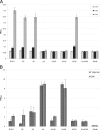
Assessment of Pel formation in various exopolysaccharide-deficient PAO1 mutants. ΔF, PAO1 ΔpelF; Δ8, PAO1 Δalg8; ΔA, PAO1 ΔpslA ΔAΔF, PAO1 ΔpslA ΔpelF; ΔAΔ8, PAO1 ΔpslA Δalg8; ΔFΔ8, PAO1 Δalg8 ΔpelF; ΔAΔFΔ8, PAO1 ΔpslA ΔpelF Δalg8. (A) Pellicle formation at air-liquid interphase when each mutant was grown in 10 ml of PI medium for 4 days as a static biofilm. (B) Congo red binding assay. All strains grown as static biofilms for 4 days were mixed with 20 mg/ml Congo red and left for 90 min while it was shaken. The biomass was sedimented, and unbound Congo red was detected at 490 nm. The percentage of Congo red left in the supernatant is shown here.
Fig. 2.
Fig. 2.
Attachment of various P. aeruginosa PAO1 strains to a solid surface. The SSA assay was used to assess the impacts of various exopolysaccharide deficiencies on attachment. ΔF, PAO1 ΔpelF; Δ8, PAO1 Δalg8; ΔA, PAO1 ΔpslA; ΔAΔF, PAO1 ΔpslA ΔpelF; ΔAΔ8, PAO1 ΔpslA Δalg8; ΔFΔ8, PAO1 Δalg8 ΔpelF; ΔAΔFΔ8, PAO1 ΔpslA ΔpelF Δalg8; media, uninoculated Pseudomonas isolation medium control. (A) Differences during early attachment phase at 2-, 4-, and 6-h time points; (B) differences between loosely and tightly attached 4-day-old biofilms (adherent biofilms after soft and vigorous washing, respectively). Values and error bars represent the averages and standard deviations, respectively, for 24 independent replicates.
Fig. 3.
Fig. 3.
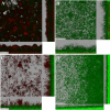
Confocal laser scanning microscopic images of P. aeruginosa biofilms grown in a continuous-culture flow cell for 4 days. The xy (upper left), xz (bottom), and yz (right) planes of each image are shown. (A) PAO1; (B) PAO1 Δalg8; (C) PAO1 ΔpelF; (D) PAO1 ΔpslA; (E) PAO1 Δalg8 ΔpelF; (F) PAO1 ΔpslA Δalg8.
Fig. 4.
Fig. 4.
DNase treatment of large amounts of extracellular DNA-producing mutants biofilms. (A) Images of PAO1 ΔpslA Δalg8 without DNase treatment; (B) images of PAO1 ΔpslA Δalg8 after DNase treatment; (C) images of PAO1 ΔpelF Δalg8 without DNase treatment; (D) images of PAO1 ΔpelF Δalg8 after DNase treatment.
Fig. 5.
Fig. 5.
Height of microcolonies and base layer of each biofilm-forming mutant measured from 3D pictures of biofilms grown for 96 h. White, height of base of the biofilm. ΔF, PAO1 ΔpelF; Δ8, PAO1 Δalg8; ΔA, PAO1 ΔpslA; ΔAΔ8, PAO1 ΔpslA Δalg8; ΔF Δ8, PAO1 Δalg8ΔpelF.

References

    1. Allesen-Holm M., et al. 2006. A characterization of DNA release in Pseudomonas aeruginosa cultures and biofilms. Mol. Microbiol. 59:1114–1128 - PubMed
    1. Campisano A., Schroeder C., Schemionek M., Overhage J., Rehm B. H. 2006. PslD is a secreted protein required for biofilm formation by Pseudomonas aeruginosa. Appl. Environ. Microbiol. 72:3066–3068 - PMC - PubMed
    1. Choi K. H., Kumar A., Schweizer H. P. 2006. A 10-min method for preparation of highly electrocompetent Pseudomonas aeruginosa cells: application for DNA fragment transfer between chromosomes and plasmid transformation. J. Microbiol. Methods 64:391–397 - PubMed
    1. Colvin K. M., et al. 2011. The Pel polysaccharide can serve a structural and protective role in the biofilm matrix of Pseudomonas aeruginosa. PLoS Pathog. 7:e1001264. - PMC - PubMed
    1. Friedman L., Kolter R. 2004. Genes involved in matrix formation in Pseudomonas aeruginosa PA14 biofilms. Mol. Microbiol. 51:675–690 - PubMed

Publication types

MeSH terms

LinkOut - more resources