. 2011 Apr;2(4):298-304.
doi: 10.18632/oncotarget.261.

Thioaptamer conjugated liposomes for tumor vasculature targeting

Affiliations

Thioaptamer conjugated liposomes for tumor vasculature targeting

Aman P Mann et al. Oncotarget. 2011 Apr.

Abstract

Recent developments in multi-functional nanoparticles offer a great potential for targeted delivery of therapeutic compounds and imaging contrast agents to specific cell types, in turn, enhancing therapeutic effect and minimizing side effects. Despite the promise, site specific delivery carriers have not been translated into clinical reality. In this study, we have developed long circulating liposomes with the outer surface decorated with thioated oligonucleotide aptamer (thioaptamer) against E-selectin (ESTA) and evaluated the targeting efficacy and PK parameters. In vitro targeting studies using Human Umbilical Cord Vein Endothelial Cell (HUVEC) demonstrated efficient and rapid uptake of the ESTA conjugated liposomes (ESTA-lip). In vivo, the intravenous administration of ESTA-lip resulted in their accumulation at the tumor vasculature of breast tumor xenografts without shortening the circulation half-life. The study presented here represents an exemplary use of thioaptamer and liposome and opens the door to testing various combinations of thioaptamer and nanocarriers that can be constructed to target multiple cancer types and tumor components for delivery of both therapeutics and imaging agent.


Figures

Figure 1
Figure 1. Physico-chemical properties of ESTA conjugated liposomes
(A) Scheme of TA conjugation to liposome. (B) Quantification of ESTA conjugation on the liposomes. Fluorescence intensity of Cy3-labeled ESTA was plotted against ESTA concentration. Using the standard curve the number of ESTA molecules on ESTA-lip was estimated based on fluorescence measurement. (C) Size and charge of the liposomes. The size of the liposomes was measured using a dynamic light scattering (DLS) and the surface charge was measured by zeta potential. (D) FTIR spectrum of the non labeled-liposomes and ESTA-lips.
Figure 2
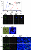
Figure 2. E-selectin dependent binding of ESTA-lip on E-selectin expressing cells
(A) Induction of E-selectin expression on the HUVEC cells. HUVEC were stimulated with TNF-α (10 ng/ml) for 4 hours. Cells were dissociated with 5mM EDTA and then incubated with FITC labeled E-selectin antibody. Surface fluorescence was measured by flow cytometry and averaged for three separate experiments. (B) ESTA-lip binding to the HUVEC expressing E-selectin. HUVEC was incubated with TNF-α (10 ng/ml) for 3 hours to induce E-selectin expression. The cells were further incubated with Cy3-ESTA-lip-FITC (10 nM) for 2 hours. All images were captured at the same exposure condition for comparison. The final images shown are representative images (at the final magnification: x600) from five random fields of at least three independent experiments. Blue, Hoechst 33342; Red, ESTA; Green, FITC-lip. (C) Immunohistochemical analysis for E-selectin expression on the vasculature of mouse bearing breast xenograft tumor derived from MDA-MB-231. ESTA-lip-Rhodamine were intravenously injected and the tumor was dissected 5 hr after the injection and frozen sections were generated to identify the localization of the liposomes by using fluorescent microscope. (D) ESTA-lip accumulation in tumor parenchyma after 48 hr after the injection. Red, lip-Rhodamine; Blue, Hoechst 33342.

Comment in

  • Is nanomedicine still promising?
    Peiris PM, Karathanasis E. Peiris PM, et al. Oncotarget. 2011 Jun;2(6):430-2. doi: 10.18632/oncotarget.295. Oncotarget. 2011. PMID: 21677362 Free PMC article. No abstract available.

References

    1. Farokhzad OC, Langer R. Impact of nanotechnology on drug delivery. ACS Nano. 2009;3(1):16–20. - PubMed
    1. Ferrari M. Cancer nanotechnology: opportunities and challenges. Nat Rev Cancer. 2005;5(3):161–171. - PubMed
    1. Qiao Y, Huang X, Nimmagadda S, Bai R, Staedtke V, Foss CA, Cheong I, Holdhoff M, Kato Y, Pomper MG, Riggins GJ, Kinzler KW, Diaz LA, Jr, Vogelstein B, Zhou S.A. robust approach to enhance tumor-selective accumulation of nanoparticles. Oncotarget. 2011;2(1-2):59–68. - PMC - PubMed
    1. Hashizume H, Baluk P, Morikawa S, McLean JW, Thurston G, Roberge S, Jain RK, McDonald DM. Openings between defective endothelial cells explain tumor vessel leakiness. Am J Pathol. 2000;156(4):1363–1380. - PMC - PubMed
    1. Less JR, Skalak TC, Sevick EM, Jain RK. Microvascular architecture in a mammary carcinoma: branching patterns and vessel dimensions. Cancer Res. 1991;51(1):265–273. - PubMed

Publication types

MeSH terms