

Profile of retinal vasculitis in a tertiary eye care center in Eastern India
- PMID: 21666315
- PMCID: PMC3129755
- DOI: 10.4103/0301-4738.81998
Profile of retinal vasculitis in a tertiary eye care center in Eastern India
Abstract
Aims: To provide a fact file on the etiology, clinical presentations and management of retinal vasculitis in Eastern India.
Materials and methods: Retrospective, record based analysis of retinal vasculitis cases in a tertiary care center in Eastern India from January 2007 to December 2009 .
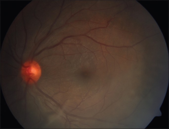
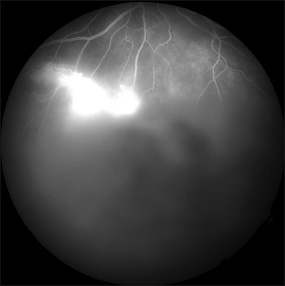
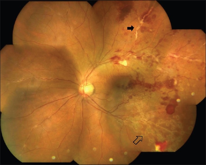
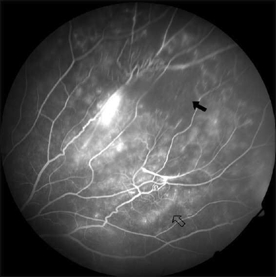
Results: One hundred and thirteen eyes of 70 patients of retinal vasculitis were included in this study. Sixty (85.7%) patients were male (mean age 33± 11.1 years) and 10 (14.3%) were female (mean age 32.4 ± 13.6 years). Vasculitis was bilateral in 43 (61.4%) and unilateral in 27 (38.6%) patients. Commonest symptoms were dimness of vision (73; 64.6%) and floaters (36; 31.9%). Vascular sheathing (82; 72.6%) and vitritis (51; 45.1%) were commonest signs. Mantoux test was positive in 21 (30%) patients but tuberculosis was confirmed in only four (5.71%) patients. Raised serum angiotensin-converting enzyme level and positive antinuclear antibody level were reported in four (5.71%) patients each. Human leukocyte antigen B5 (HLA B5) marker was present in one (1.4%) patient. However, none of the total 70 patients were found to have a conclusively proven systemic disease attributable as the cause of retinal vasculitis. Oral corticosteroid (60; 85.7%) was the mainstay of treatment. Forty-eight (42.5%) eyes maintained their initial visual acuity and 43 (38%) gained one or more line at mean follow-up of 16.6± 6.3 months.
Conclusion: Retinal vasculitis cases had similar clinical presentations and common treatment plan. There was no systemic disease association with vasculitis warranting a careful approach in prescribing investigations.
Conflict of interest statement
Figures
Comment in
-
Ocular manifestations of the antineutrophil cytoplasmic antibody and antiphospholipid syndromes.Indian J Ophthalmol. 2013 Jul;61(7):365-6. doi: 10.4103/0301-4738.111127. Indian J Ophthalmol. 2013. PMID: 23619483 Free PMC article. No abstract available.
References
-
- Walton RC, Ashmore ED. Retinal vasculitis. Curr Opin Ophthalmol. 2003;14:413–9. - PubMed
-
- Abu El-Asrar AM, Herbort CP, Tabbara KF. Retinal vasculitis. Ocul Immunol Inflamm. 2005;13:415–33. - PubMed
-
- Perez VL, Chavala SH, Ahmed M, Chu D, Zafirakis P, Baltatzis S, et al. Ocular manifestations and concepts of systemic vasculitides. Surv Ophthalmol. 2004;49:399–418. - PubMed
MeSH terms
Substances
LinkOut - more resources
Full Text Sources
Medical
Research Materials
