. 2011 Jun 12;474(7352):472-6.
doi: 10.1038/nature10178.

Structural insight into brassinosteroid perception by BRI1

Affiliations

Structural insight into brassinosteroid perception by BRI1

Ji She et al. Nature. .

Abstract

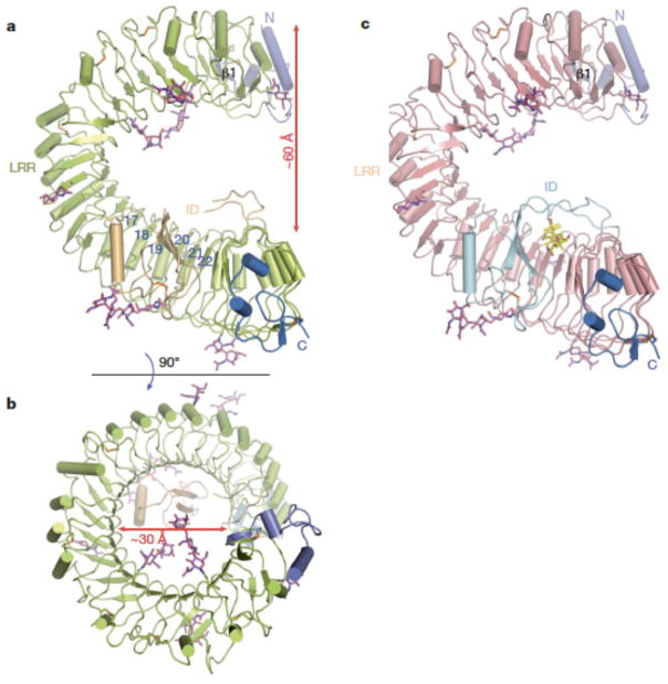
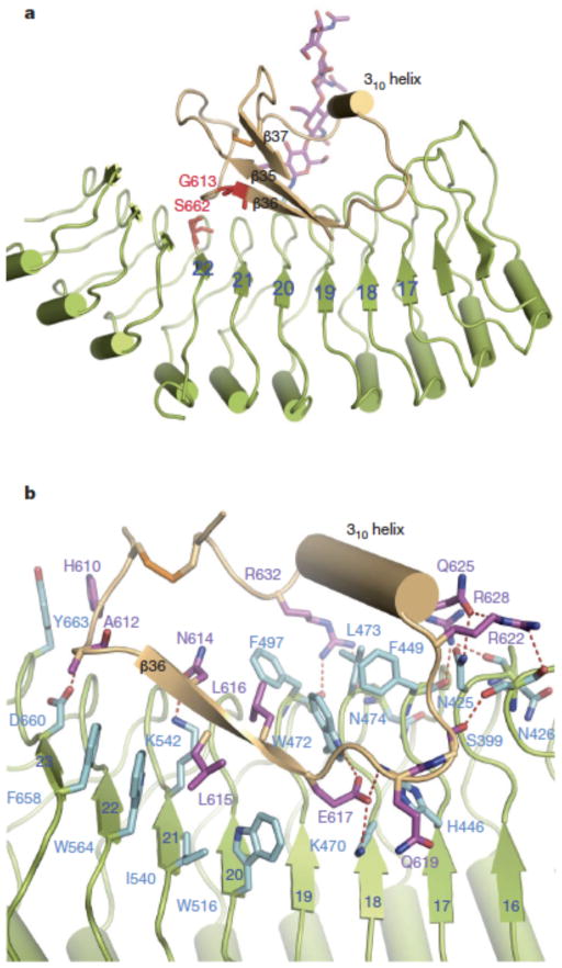
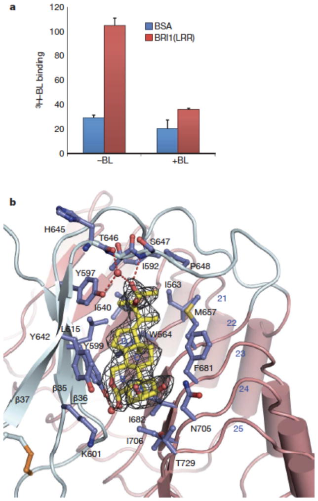
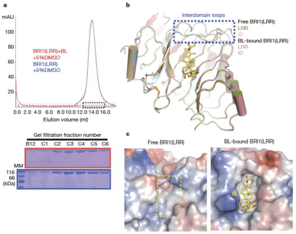
Brassinosteroids are essential phytohormones that have crucial roles in plant growth and development. Perception of brassinosteroids requires an active complex of BRASSINOSTEROID-INSENSITIVE 1 (BRI1) and BRI1-ASSOCIATED KINASE 1 (BAK1). Recognized by the extracellular leucine-rich repeat (LRR) domain of BRI1, brassinosteroids induce a phosphorylation-mediated cascade to regulate gene expression. Here we present the crystal structures of BRI1(LRR) in free and brassinolide-bound forms. BRI1(LRR) exists as a monomer in crystals and solution independent of brassinolide. It comprises a helical solenoid structure that accommodates a separate insertion domain at its concave surface. Sandwiched between them, brassinolide binds to a hydrophobicity-dominating surface groove on BRI1(LRR). Brassinolide recognition by BRI1(LRR) is through an induced-fit mechanism involving stabilization of two interdomain loops that creates a pronounced non-polar surface groove for the hormone binding. Together, our results define the molecular mechanisms by which BRI1 recognizes brassinosteroids and provide insight into brassinosteroid-induced BRI1 activation.


Conflict of interest statement

The authors declare no competing financial interests.

Figures

Fig. 1
Fig. 1. BRI1-LRR has a helical solenoid structure
a. and (b) are overall structures of BL-free BRI1-LRR shown in two different orientations. The N-linked sugars (N-acetylglucosamines) are shown in magenta stick. Colored in orange are disulfide bonds. The N- and C-terminal cap is shown in slate and marine respectively. The blue numbers in (a) indicate the positions of LRRs. The β-strand (β1) from the N-terminal cap is labeled. c. Overall structure of BL-bound BRI1-LRR with the same orientation as (a). BL molecule is shown in stick and colored in yellow.
Fig. 2
Fig. 2. Interaction of the ID with LRRs
a. Cartoon representation of BRI1-LRR highlighting interaction of the ID with the concave surface. The positions of two genetic mutants are shown in red and stick. b. Detailed interactions of the ID with LRRs. The side chains from the ID are shown and labeled in purple, and those from LRRs in cyan. Red dashed lines represent hydrogen bonds.
Fig. 3
Fig. 3. BL binds a hydrophobic groove between the ID and the inner surface of LRRs
a. [3H]-BL binding activity of BRI1-LRR. About 1 mg ml−1 BRI1-LRR-His (red bar) or BSA as control (blue bar) was incubated with 20 nM [3H]-BL in the absence (−BL) or presence (+BL) of 20 μM unlabelled BL. BRI1-bound [3H]-BL was recovered using nickel beads and quantified by scintillation counting. Data represent the average of triplicate assays and error bars are standard deviations. b. Detailed interactions between BL and BRI1-LRR. Shown in mesh is omit electron density (2.5 δ) around BL. The ID and LRRs are colored in slate and salmon, respectively. The side chains from both the ID and LRRs are shown in slate. Red spheres represent oxygen atoms of water molecules. The three β–strands from the ID are labeled.
Fig. 4
Fig. 4. BL induces stabilization of two inter-domain loops but no dimerization of BRI1-LRR
a. BL has no effect on the oligomeric status of BRI1-LRR in solution. Shown on the top is superposition of the gel filtration chromatograms of BRI1-LRR in the absence (blue) and presence (red) of BL. The vertical and horizontal axes represent UV absorbance (λ=280 nm) and elution volume respectively. Peak fractions are highlighted within the dashed square. The apparent molecular weight of BRI1-LRR was 109.4 kD in the presence or absence of BL, higher than the theoretical BRI1-LRR monomer (83 kD) likely due to existence of multiple glycosylation sites in BRI1-LRR. MM: molecular mass marker. Bottom: Coomassie blue staining of the peak fractions shown on the top following SDS-PAGE. b. BL binding induces stabilization of two inter-domain loops. Structural superimposition of the free and BL-bound BRI1-LRR around the BL-binding site. c. BL binding generates a striking hydrophobic surface groove on BRI1-LRR. Shown on the left and right are the electrostatic surfaces of free BRI1-LRR and BL-bound BRI1-LRR (shown in the same orientation) around the BL-binding site respectively. The area highlighted with the yellow dashed square on the left panel is the BL-binding site. White, blue and red indicate neutral, positive and negative surfaces respectively.

References

    1. Clouse SD, Sasse JM. BRASSINOSTEROIDS: Essential Regulators of Plant Growth and Development. Annu Rev Plant Physiol Plant Mol Biol. 1998;49:427–451. - PubMed
    1. Vert G, Nemhauser JL, Geldner N, Hong F, Chory J. Molecular mechanisms of steroid hormone signaling in plants. Annu Rev Cell Dev Biol. 2005;21:177–201. - PubMed
    1. Li J, Chory J. A putative leucine-rich repeat receptor kinase involved in brassinosteroid signal transduction. Cell. 1997;90:929–38. - PubMed
    1. Li J, et al. BAK1, an Arabidopsis LRR receptor-like protein kinase, interacts with BRI1 and modulates brassinosteroid signaling. Cell. 2002;110:213–22. - PubMed
    1. Nam KH, Li J. BRI1/BAK1, a receptor kinase pair mediating brassinosteroid signaling. Cell. 2002;110:203–12. - PubMed

Publication types

MeSH terms