. 2011 Dec 8;30(49):4901-9.
doi: 10.1038/onc.2011.187. Epub 2011 Jun 13.

C/EBP-δ regulates VEGF-C autocrine signaling in lymphangiogenesis and metastasis of lung cancer through HIF-1α

Affiliations

C/EBP-δ regulates VEGF-C autocrine signaling in lymphangiogenesis and metastasis of lung cancer through HIF-1α

Y Min et al. Oncogene. .

Abstract

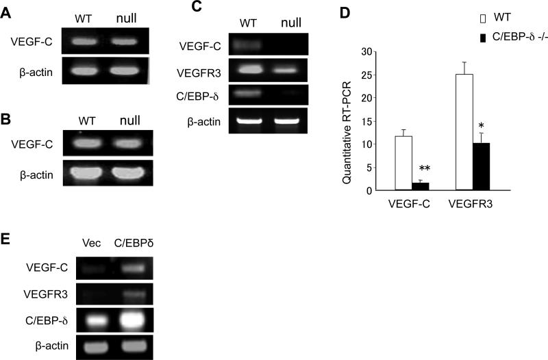
CCAAT/enhancer-binding protein-δ (C/EBP-δ), a transcription factor, is elevated in carcinoma compared with that in normal tissue. This study reports a novel function of C/EBP-δ in lymphangiogenesis and tumor metastasis. Genetic deletion of C/EBP-δ in mice resulted in a significant reduction of lymphangiogenesis and pulmonary metastases, with a dramatic reduction of vascular endothelial growth factor-C (VEGF-C) and its cognate receptor VEGF receptor-3 (VEGFR3) in lymphatic endothelial cells (LECs). By contrast, no difference of VEGF-C in tumor tissues and bone marrow was observed between null and wild-type mice. Consistently, forced expression of C/EBP-δ increased VEGF-C and VEGFR3 expression in cultured LECs. These findings suggest a specific and important role of C/EBP-δ in the regulation of VEGFR3 signaling in LECs. Furthermore, expression of C/EBP-δ in cultured LECs significantly increased cell motility, and knockdown of C/EBP-δ inhibited cell motility and lymphatic vascular network formation in vitro. Forced expression of VEGF-C, but not recombinant VEGF-C, rescued the knockdown of C/EBP-δ-induced cell apoptosis, indicative of autonomous VEGF-C autocrine signaling essential for LEC survival. Moreover, hypoxia induces C/EBP-δ expression and C/EBP-δ regulates HIF-1α expression. Blocking HIF-1α activity totally blocked CEBP-δ-induced VEGF-C and VEGFR3 expression in LECs. Together, these findings uncover a new function of CEBP-δ in lymphangiogenesis through regulation of VEGFR3 signaling in LECs.


Figures

Figure 1
Figure 1
Inactivation of C/EBP-δ in mice inhibits tumor lymphangiogenesis and pulmonary metastasis of lung cancer. 1×10⁵ 3LL cells were injected via tail vein into 6-week old female wild type and C/EBP-δ null mice. Fourteen days later, lungs were harvested and imaged (Panel A). Representative images are shown. Arrows point to tumor nodules. Tumor metastasis was quantified by counting the number of lung surface metastases (Panel B) and measuring the mass of the lungs (Panel C). Data are expressed as mean ± SD. n=10 mice per group, *p<0.05. Tumor nodules in lungs were subjected to immunohistochemical analysis for Lyve-1, a marker for lymph endothelium (Panel D). 200X Magnification. The number of Lyve-1 positive lymphatic vessels was counted from 10 randomly selected high power fields under microscopy (Panel E). Data are expressed as mean ± SD. *p<0.05.
Figure 2
Figure 2
C/EBP-δ regulates VEGF-C and VEGFR3 expression in lymphatic endothelial cells. Total RNA was isolated from tumor tissues (Panel A) and bone marrow (Panel B) of wild type and C/EBP-δ null mice, and subjected to semi-quantitative RT-PCR for VEGF-C. Pulmonary lymphatic microvascular endothelial cells were isolated from age and sex matched wild type and C/EBP-δ null mice (pooled from 5 mice per group), and purified with flow cytometry cell sorting using anti Lyve-1-PE antibodies. Total RNA isolated from the murine lymphatic endothelial cells was subjected to semi-quantitative RT-PCR (Panel C) and real-time PCR (Panel D) for VEGF-C and VEGFR-3. *p<0.05, **p<0.01. HMVEC-LLy cells were transfected with either empty vector or C/EBP-δ expression vector for 48 hours. Total RNA was isolated and subjected to semi-quantitative RT-PCR for genes indicated (Panel E). Each experiment was repeated at least three times. Representative images are shown.
Figure 3
Figure 3
C/EBP-δ regulates lymphangiogenesis in vitro. HMVEC-LLy cells were transfected with a C/EBP-δ expression vector (C/EBP-δ, Panel A), or knockdown using specific shRNA construct for C/EBP-δ (K’C/EBP-δ, Panel B). Empty vector and non-specific shRNA were used as controls. Expression of C/EBP-δ in lymphatic endothelial cells was determined by semi quantitative RT-PCR. Lymphatic endothelial cell migration was measured in forced expression and knockdown cells using Transwell assays in response to 50 ng/ml of VEGF-C stimulation. Migrated cells were counted in 10 randomly selected 200X high power fields under microscopy after a 5-hour incubation (Panel C and Panel D). **p<0.01. Lymphatic vascular network formation was assessed using the Matrigel assay. Vascular network formation in Matrigel was imaged under microscopy 24 hrs after cell plating in C/EBP-δ forced expression cells (Panel E) and C/EBP-δ knockdown cells (Panel G). Vascular cross points were counted from 10 randomly selected high power fields under microscopy (Panel F and Panel H). The data were collected from three independent experiments performed in triplicate, and expressed as mean ± SD. **p<0.01.
Figure 4
Figure 4
C/EBP-δ regulates VEGF-C autocrine signaling in lymphatic endothelial cell survival. HMVEC-LLy cells were transfected with either shRNA vector for C/EBP-δ (K’C/EBP-δ) or control vector for 48 hours, followed by incubation with PI and antibody against Annexin V. Cell apoptosis was assessed by flow cytometry (Panel A). C/EBP-δ knockdown HMVEC-LLy cells were either incubated with 50 ng/ml of recombinant VEGF-C or co-transfected with a VEGFC expression vector. Cell apoptosis was assessed by PI and Annexin V staining and flow cytometry (Panel B). Representative images were shown. Live cells were quantitated in each group (Panel C). Each experiment was done in duplicate and repeated three times. Data are expressed as mean ± SE. *p<0.01 vs control, **p<0.01 vs K’C/EBP-δ.
Figure 5
Figure 5
C/EBP-δ regulates VEGF-C and VEGFR3 production in lymphatic endothelial cells through HIF-1α. Purified murine pulmonary LECs from wild type and C/EBP-δ null mice were subjected to semi-quantitative RT-PCR for HIF-1α and C/EBP-δ expression (Panel A). HMVEC-LLy cells were cultured under normoxia (20% O2) or hypoxia (1% O2) for 24 hours. Semi-quantitative RT-PCR was performed to measure C/EBP-δ (Panel B). HMVEC-LLy cells were transfected with either empty vector or C/EBP-δ expression vector for 24 hours. The cells were then cultured under normoxia (20% O2) or hypoxia (1% O2) for another 48 hours. Transcript levels of VEGF-C, VEGFR3, HIF-1α and C/EBP-δ were measured by semi-quantitative RT-PCR (Panel C). HMVEC-LLy cells were transfected with either control shRNA or shRNA for C/EBP-δ for 24 hours. The cells were then cultured under either normoxia (20% O2) or hypoxia (1% O2) for another 48 hours. Transcript levels of VEGF-C, VEGFR3, HIF-1α and C/EBP-δ were measured by semi-quantitative RT-PCR (Panel D). HMVEC-LLy cells were transfected with either empty vector or C/EBP-δ expression vector for 24 hours. The cells were treated with or without HIF-1α inhibitor (5 μM of geldanamycin) for another 48 hours under hypoxic conditions. Transcript levels of VEGF-C, VEGFR3, HIF-1α and C/EBP-δ were measured by semi-quantitative RT-PCR (Panel E). Each experiment was repeated three times. Representative images are shown.
Figure 6
Figure 6
Schematic diagram of C/EBP-δ in regulating VEGF-C signaling.

References

    1. Akagi K, Ikeda Y, Miyazaki M, Abe T, Kinoshita J, Maehara Y, et al. Vascular endothelial growth factor-C (VEGF-C) expression in human colorectal cancer tissues. Br J Cancer. 2000;83:887–891. - PMC - PubMed
    1. Alitalo K, Carmeliet P. Molecular mechanisms of lymphangiogenesis in health and disease. Cancer Cell. 2002;1:219–227. - PubMed
    1. Alitalo K, Tammela T, Petrova TV. Lymphangiogenesis in development and human disease. Nature. 2005;438:946–953. - PubMed
    1. Dumont DJ, Jussila L, Taipale J, Lymboussaki A, Mustonen T, Pajusola K, et al. Cardiovascular failure in mouse embryos deficient in VEGF receptor-3. Science. 1998;282:946–949. - PubMed
    1. He Y, Kozaki K, Karpanen T, Koshikawa K, Yla-Herttuala S, Takahashi T, et al. Suppression of tumor lymphangiogenesis and lymph node metastasis by blocking vascular endothelial growth factor receptor 3 signaling. J Natl Cancer Inst. 2002;94:819–825. - PubMed

Publication types

MeSH terms

Substances