

Immune recognition and rejection of allogeneic skin grafts
- PMID: 21668313
- PMCID: PMC3738014
- DOI: 10.2217/imt.11.2
Immune recognition and rejection of allogeneic skin grafts
Abstract
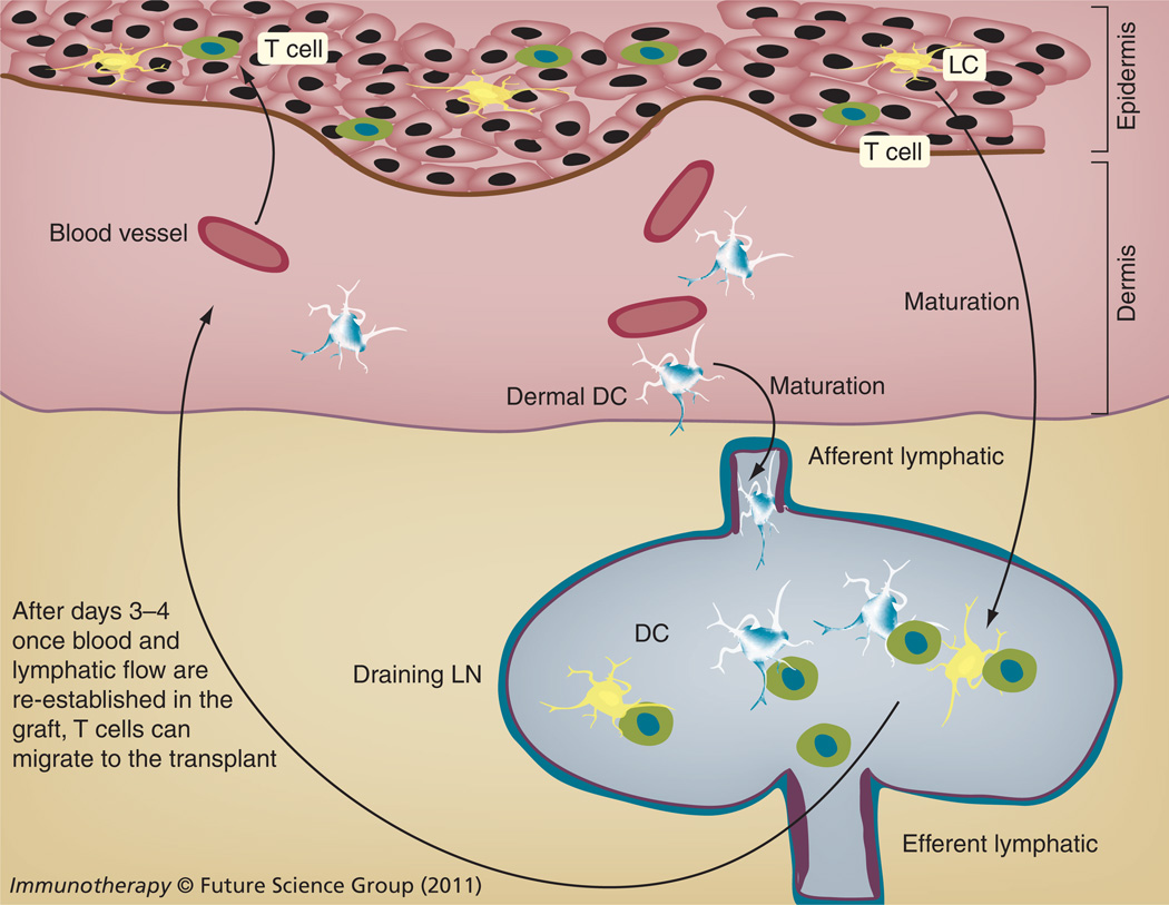
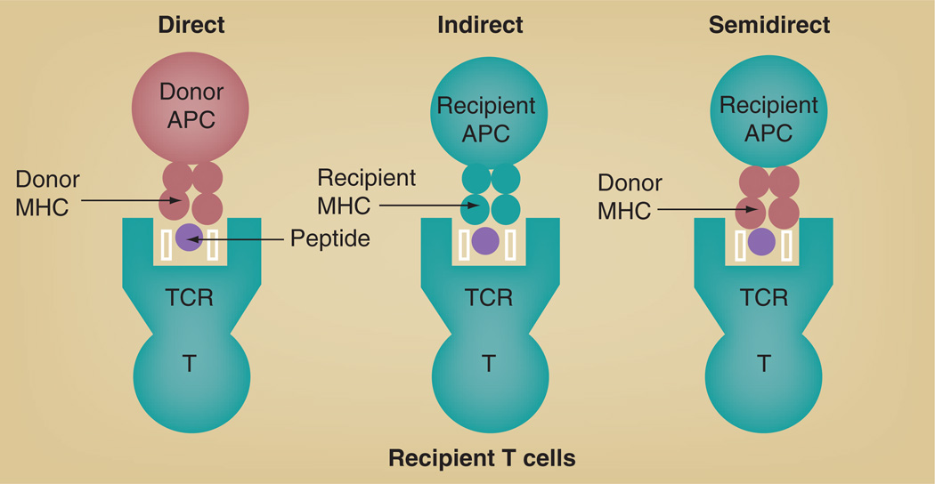
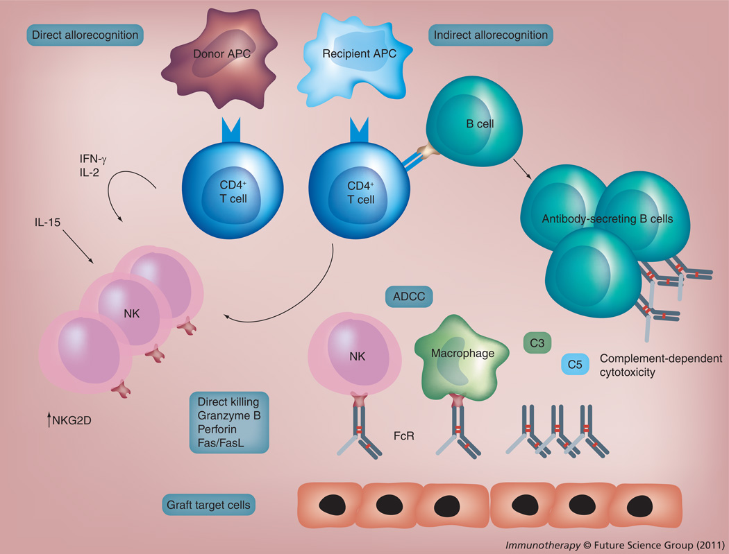
The transplantation of allogeneic skin grafts is associated with a potent inflammatory immune response leading to the destruction of donor cells and the rejection of the graft. Shortly after transplantation, skin dendritic cells (DCs) migrate out of the graft through lymphatic vessels and infiltrate the recipient's draining lymph nodes where they present donor antigens via two mechanisms: the direct pathway, in which T cells recognize intact donor MHC antigens on donor DCs; and the indirect pathway, involving T-cell recognition of donor peptides bound to self-MHC molecules on recipient DCs. Some recent studies have suggested that T cells can become activated via recognition of donor MHC molecules transferred on recipient antigen-presenting cells (semidirect pathway). Activation of T cells via direct or indirect allorecognition is sufficient to trigger acute rejection of allogeneic skin grafts. In addition, allospecific antibodies contribute to the rejection process either by killing allogeneic targets in a complement-dependent fashion or by opsonizing donor cells and forming immune complexes. Finally, several studies demonstrate that NK cells, activated due to missing self-MHC class I molecules on allogeneic cells, are involved in allogeneic skin graft rejection via direct killing of donor cells and through the production of proinflammatory cytokines including IFN-γ and TNF-α.
Conflict of interest statement
The authors have no other relevant affliations or financial involvement with any organization or entity with a fnancial interest in or financial conflict with the subject matter or materials discussed in the manuscript apart from those disclosed.
No writing assistance was utilized in the production of this manuscript.
Figures
References
-
- Billingham RE, Medawar PD. The technique of free skin grafting in mammals. J. Exp. Biol. 1951;28:385.
-
- Billingham RE, Brent L, Medawar PB. Activity acquired tolerance of foreign cells. Nature. 1953;172(4379):603–606. - PubMed
-
- Rosenberg AS, Singer A. Cellular basis of skin allograft rejection: an in vivo model of immune-mediated tissue destruction. Annu. Rev. Immunol. 1992;10:333–358. - PubMed
-
- Valladeau J, Ravel O, Dezutter-Dambuyant C, et al. Langerin, a novel c-type lectin specific to langerhans cells, is an endocytic receptor that induces the formation of Birbeck granules. Immunity. 2000;12(1):71–81. - PubMed
Publication types
MeSH terms
Grants and funding
LinkOut - more resources
Full Text Sources
Other Literature Sources
Research Materials