

Developmental changes in the sleep electroencephalogram of adolescent boys and girls
- PMID: 21668552
- PMCID: PMC3987854
- DOI: 10.1111/j.1365-2869.2011.00930.x
Developmental changes in the sleep electroencephalogram of adolescent boys and girls
Abstract
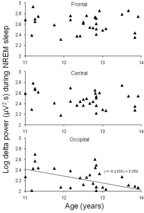
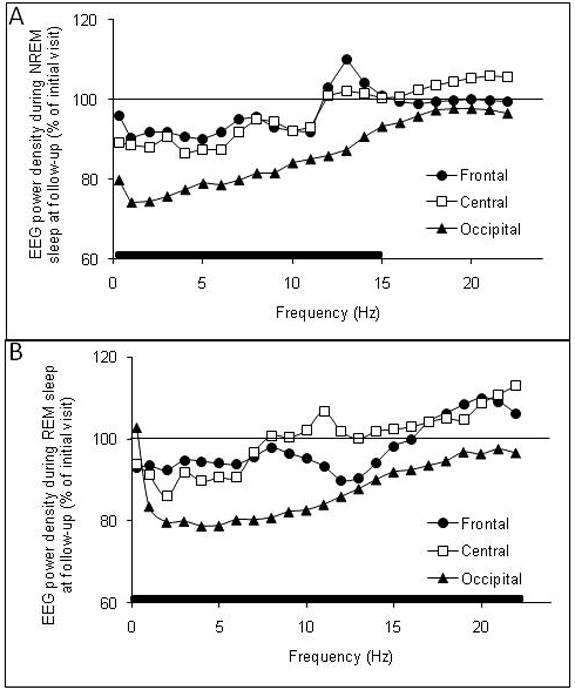
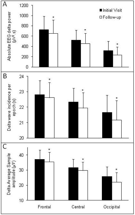
The sleep electroencephalogram (EEG) changes across adolescence; however, there are conflicting data as to whether EEG changes are regionally specific, are evident in non-rapid eye movement (NREM) and rapid eye movement (REM) sleep, and whether there are sex differences. The present study seeks to resolve some of these issues in a combined cross-sectional and longitudinal analysis of sleep EEG in adolescents. Thirty-three healthy adolescents (18 boys, 15 girls; 11-14 years) were studied on two occasions 6-8 months apart. Cross-sectional analysis of data from the initial visit revealed significantly less slow-wave sleep, delta (0.3 to <4 Hz) and theta (4 to <8 Hz) power in both NREM and REM sleep with advancing age. The age-delta power relationship was significant at the occipital site, with age accounting for 26% of the variance. Longitudinal analysis revealed that NREM delta power declined significantly from the initial to follow-up visit, in association with declining delta amplitude and incidence (P < 0.01), with the effect being greatest at the occipital site. REM delta power also declined over time in association with reduced amplitude (P < 0.01). There were longitudinal reductions in theta, alpha and sigma power in NREM and REM sleep evident at the occipital site at follow-up (P < 0.01). No sex differences were apparent in the pattern of change with age for NREM or REM sleep. Declines in sleep EEG spectral power occur across adolescence in both boys and girls, particularly in the occipital derivation, and are not state-specific, occurring in both NREM and REM sleep.
© 2011 European Sleep Research Society.
Conflict of interest statement
Disclosures: Other authors have no conflict of interest to report.
Figures
References
-
- Buchmann A, Ringli M, Kurth S, et al. EEG sleep slow-wave activity as a mirror of cortical maturation. Cereb Cortex. 2011;21:607–15. - PubMed
-
- Carskadon MA, Acebo C. A self-administered rating scale for pubertal development. J Adolesc Health. 1993;14:190–5. - PubMed
-
- Carskadon MA, Orav EJ, Dement WC. Evolution of sleep and daytime sleepiness in adolescents. New York: Raven Press; 1983.
Publication types
MeSH terms
Grants and funding
LinkOut - more resources
Full Text Sources
Medical
