

Oligodendrocyte vulnerability following traumatic brain injury in rats
- PMID: 21669255
- PMCID: PMC3523350
- DOI: 10.1016/j.neulet.2011.05.056
Oligodendrocyte vulnerability following traumatic brain injury in rats
Erratum in
- Neurosci Lett. 2012 May 10;516(1):166. de Rivero Vaccari, Juan [corrected to de Rivero Vaccari, Juan Pablo]
Abstract
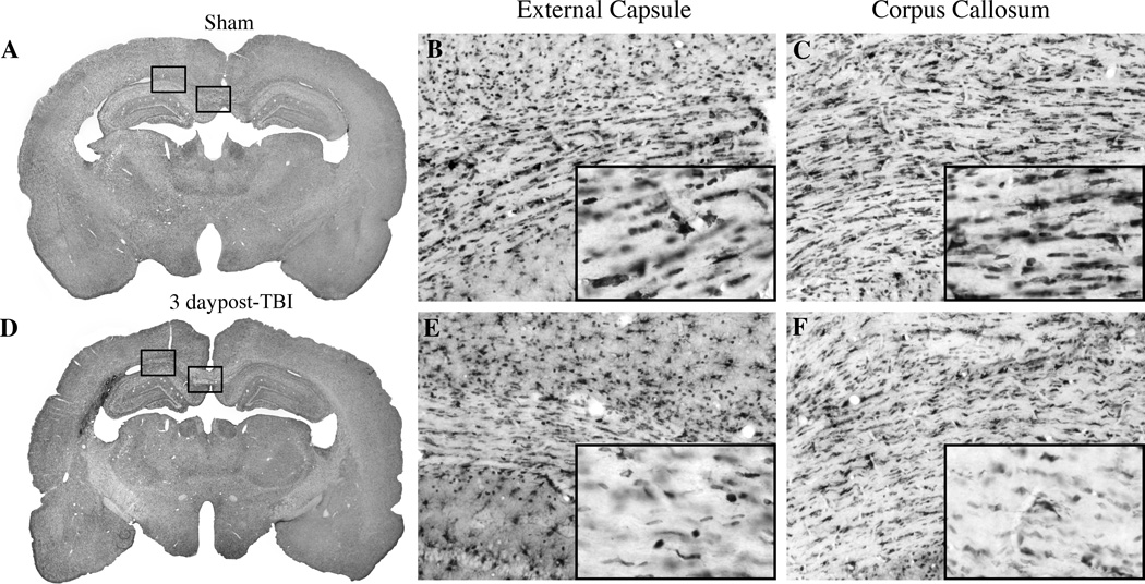
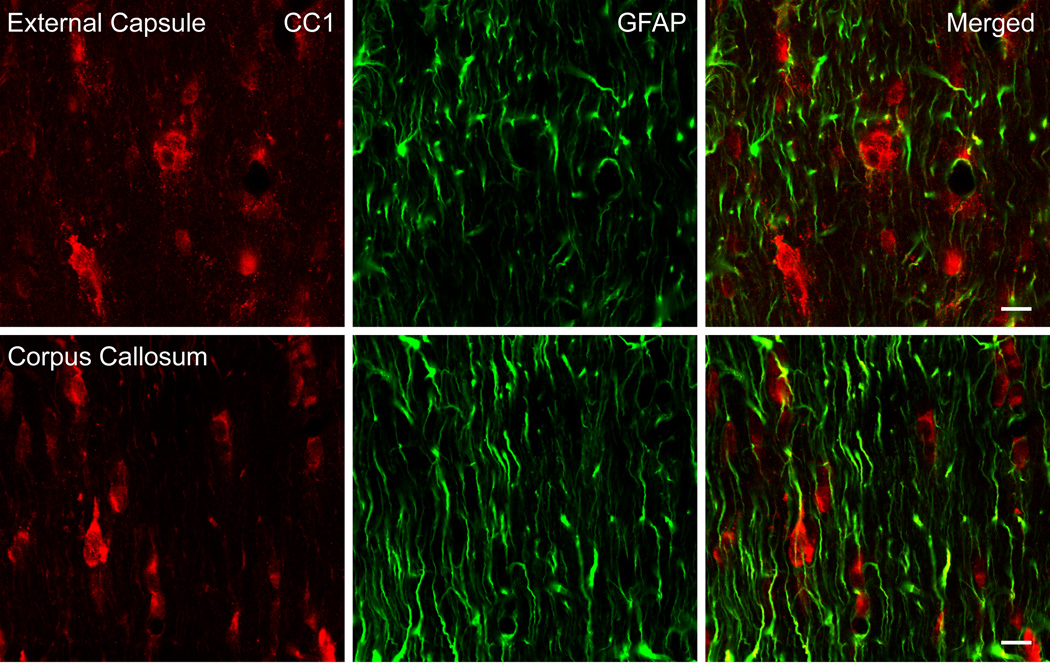
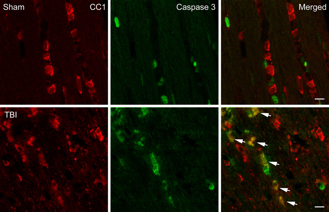
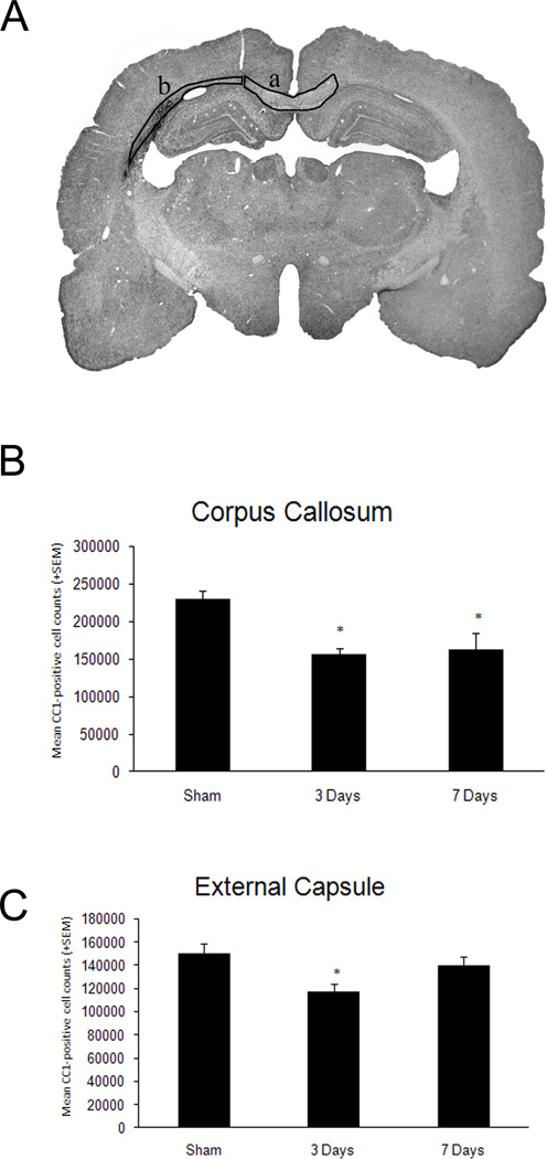
Experimental and clinical findings demonstrate that traumatic brain injury (TBI) results in injury to both gray and white matter structures. The purpose of this study was to document patterns of oligodendrocyte vulnerability to TBI. Sprague Dawley rats underwent sham operated procedures or moderate fluid percussion brain injury. Quantitative immunohistochemical analysis was performed on animals perfusion-fixed at 3 (n=9) or 7 (n=9) days post-surgery. Within the ipsilateral external capsule and corpus callosum, numbers of APC-CC1 immunoreactive oligodendrocytes were significantly decreased at 3 or 7 days post-TBI compared to sham rats (p<0.03). At both posttraumatic survival periods, double-labeling studies indicated that oligodendrocytes showed increased Caspase 3 activation compared to sham. These data demonstrate regional patterns of oligodendrocyte vulnerability after TBI and that oligodendrocyte cell loss may be due to Caspase 3-mediated cell death mechanisms. Further studies are needed to test therapeutic interventions that prevent trauma-induced oligodendrocyte cell death, subsequent demyelination and circuit dysfunction.
Copyright © 2011 Elsevier Ireland Ltd. All rights reserved.
Figures
References
-
- Bhat RV, Axt KJ, Fosnaugh JS, Smith KJ, Johnson KA, Hill DE, Kinzler KW, Baraban JM. Expression of the APC tumor suppressor protein in oligodendroglia. Glia. 1996;17:169–174. - PubMed
-
- Bramlett HM, Dietrich WD. Pathophysiology of cerebral ischemia and brain trauma: similarities and differences. J Cereb Blood Flow Metab. 2004;24:133–150. - PubMed
-
- Bramlett HM, Dietrich WD. Progressive damage after brain and spinal cord injury: pathomechanisms and treatment strategies. Prog Brain Res. 2007;161:125–141. - PubMed
-
- Bramlett HM, Dietrich WD. Quantitative structural changes in white and gray matter 1 year following traumatic brain injury in rats. Acta Neuropathol (Berl) 2002;103:607–614. - PubMed
-
- Bramlett HM, Kraydieh S, Green EJ, Dietrich WD. Temporal and regional patterns of axonal damage following traumatic brain injury: a beta-amyloid precursor protein immunocytochemical study in rats. J Neuropathol Exp Neurol. 1997;56:1132–1141. - PubMed
Publication types
MeSH terms
Substances
Grants and funding
LinkOut - more resources
Full Text Sources
Research Materials
