. 2011 Aug 1;71(15):5144-53.
doi: 10.1158/0008-5472.CAN-11-0425. Epub 2011 Jun 13.

Inhibition of miR-193a expression by Max and RXRα activates K-Ras and PLAU to mediate distinct aspects of cellular transformation

Affiliations

Inhibition of miR-193a expression by Max and RXRα activates K-Ras and PLAU to mediate distinct aspects of cellular transformation

Dimitrios Iliopoulos et al. Cancer Res. .

Abstract

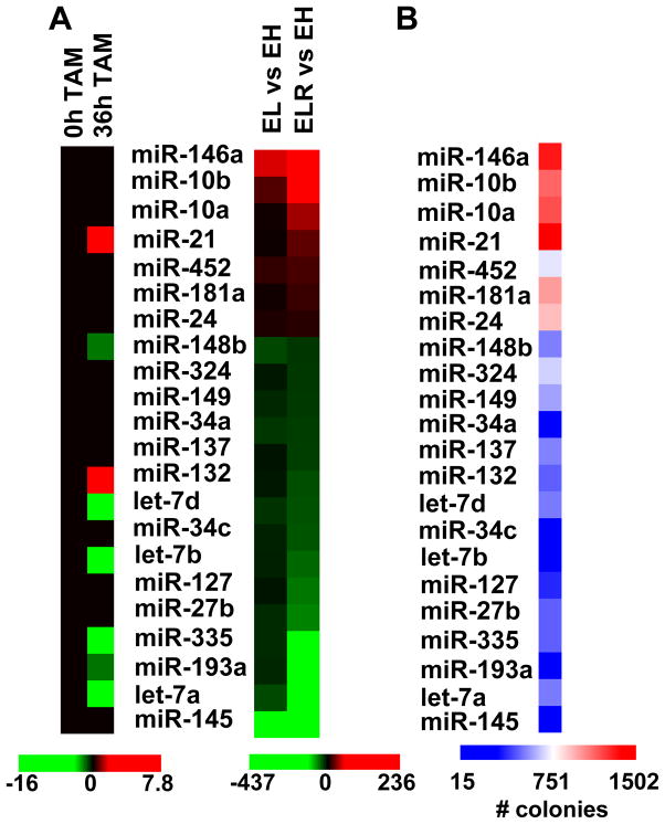
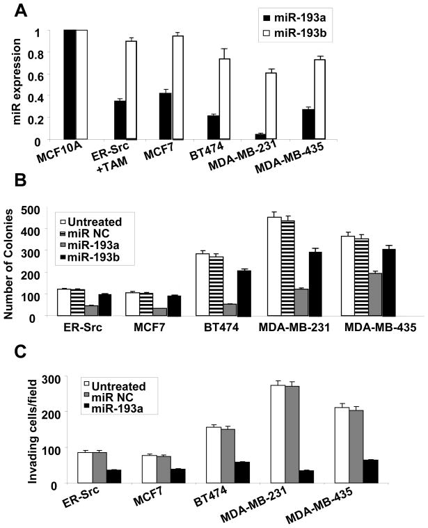
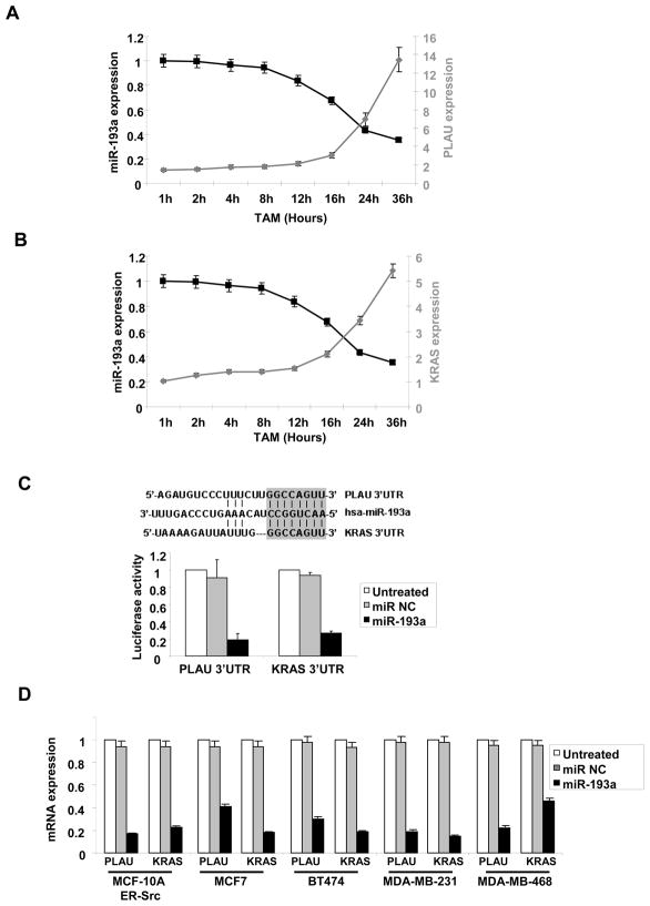
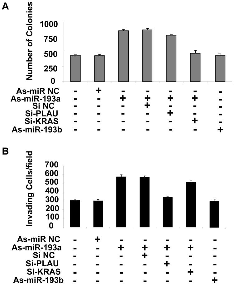
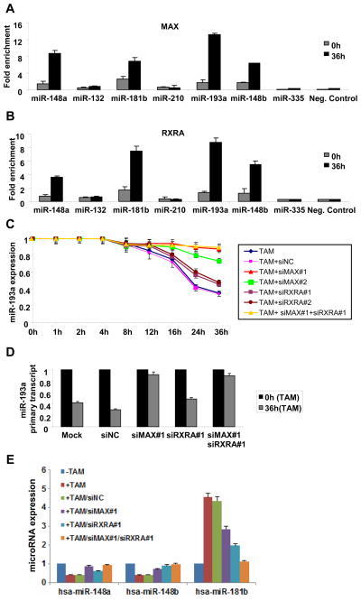
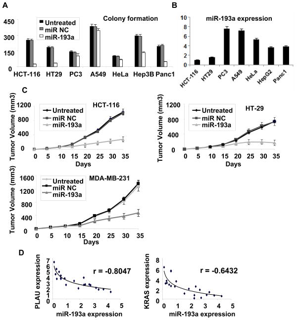
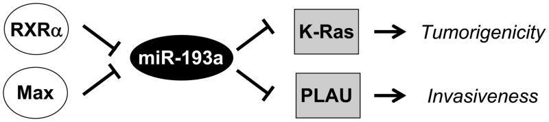
MicroRNA profiling in isogenic models of cellular transformation involving either breast epithelial cells or fibroblasts reveals that expression of miR-193a is lower in transformed cells than in nontransformed cells. The transcription factors Max and RXRα bind directly to the miR-193a promoter and inhibit miR-193a expression during transformation. miR-193a inhibits cellular transformation by directly targeting the 3' untranslated regions of PLAU and K-Ras. Interestingly, miR-193a controls anchorage-independent growth in soft agar through K-Ras, whereas it affects invasive growth through PLAU. miR-193a overexpression inhibits the tumorigenicity of developmentally diverse but not all cancer cell types, and it inhibits tumor growth in colon- and breast-derived xenografts. Finally, expression of miR-193a is inversely correlated with PLAU and K-Ras in human colon adenocarcinomas. Thus, a pathway in which Max and RXRα inhibit miR-193a expression, thereby activating the PLAU and K-Ras oncogenes is important for distinct aspects of cellular transformation, as well as tumor growth and colon (and perhaps other types of) cancer.


Figures

Figure 1
Figure 1
Identification of miRNAs that are differentially expressed and show tumor suppressor activity in the isogenic fibroblast model. A, Heat map representation of differentially expressed miRNAs in EH (immortalized), EL (pre-disposed), and ELR (transformed) fibroblasts are shown on the right. Expression of these miRNAs in non-transformed or transformed (tamoxifen-treated for 36h) breast epithelial (MCF-10A-ER-Src) cells, as determined previously (7), are shown on the left. B, Heatmap representation of tumorigenicity (number of colonies in soft agar) of transformed (ELR) fibroblasts after transfection of miRNAs. All miRNAs that significantly increase (> 1100 colonies) or decrease (< 300 colonies) tumorigenicity with respect the ~800 colonies generated by cells transfected by control miRNAs) and/or are differentially expressed are included in the heat maps. The values represent the mean of three independent experiments.
Figure 2
Figure 2
MiR-193a and miR-193b expression levels and effects on tumorigenicity and invasive growth in breast cancer cell lines. A, Relative MiR-193a and miR-193b RNA levels in the indicated cell lines. Values for miR-193a and miR-193b in the non-transformed MCF-10A cells are arbitrarily set to 1. Based on comparable PCR amplification efficiencies determined experimentally, miR-193b appears to be approximately 3-fold more abundant than miR-193a in MCF-10A cells. B, Colony formation in soft agar of the indicated breast cancer cell lines transfected with miR-193a, miR-193b, or control miRNAs. C, Invasive growth (invading cell/field after wounding) of the indicated breast cancer cell lines transfected with miR-193a control miRNAs.
Figure 3
Figure 3
MiR-193a targets PLAU and K-Ras. A, Relative expression levels of PLAU and miR-193a during cellular transformation of ER-Src cells at the indicated times points after treatment with tamoxifen. B, Relative expression levels of K-Ras and miR-193a during cellular transformation of ER-Src cells at the indicated times points after treatment with tamoxifen. C, Sequence complementarity (vertical lines) between miR-193a and the 3′ UTRs of PLAU and K-Ras, with the 8 nt seed sequence shown as a gray box. Luciferase activity of reporters containing the 3′UTR of PLAU or K-Ras 24h after transfection with miR-193a or miR negative control. D, PLAU or K-Ras mRNA levels in the indicated breast cancer cell lines transfected with miR-193a or control miRNA.
Figure 4
Figure 4
Combined effects of miR-193a and PLAU or K-Ras tumorigenicity and invasive growth. Tamoxifen-treated ER-Src cells transfected with the indicated oligonucleotides - anti-sense (As) against miR-193a or miR-193b, siRNAs against PLAU and K-Ras, and negative controls – were assayed for A, number of colonies in soft agar and B, invasive growth.
Figure 5
Figure 5
Max and RXRα bind the miR-193a and other promoters and inhibit their expression. A, MAX and B, RXRα occupancy (fold enrichment) at the miR-148a, miR-132, miR-181b, miR-210, miR-193a, miR-148b and miR-335 loci as determined by chromatin immunoprecipitation of ER-Src cells that were or were not treated with TAM. C, miR-193a expression in ER-Src cells treated with tamoxifen for the indicated amount of time in the presence or absence of two siRNAs against MAX and two siRNAs against RXRA or their combination. D, Expression levels of miR-193a primary transcript in non-transformed and transformed ER-Src cells treated with siRNA control or siRNA against Max and/or RXRα. E, Expression of the indicated miRNAs in ER-Src cells treated with tamoxifen for 36h in the presence or absence of siRNAs against MAX, RXRα, or their combination.
Figure 6
Figure 6
Importance of MiR-193a in other cancer cell types, mouse xenografts, and human cancer patients. A, Soft agar colony formation in lung (A549), hepatocellular (Hep3B), pancreatic (Panc-1), prostate (PC3), cervical (HeLa) and two colon (HT29, HCT-116) cancer cell lines treated with miR-193a or control miRNA. B, Expression of miR-193a in the indicated cancer cell lines. C, Tumor growth (mean ± SD) of colon (HT29 and HCT-116) and breast (MDA-MB-231) cancer cells after intraperitoneal treatment with miR-193a or control miRNA (days 10, 15, 20 after the initial injection of cancer cells). D, miR-193a, PLAU and K-Ras expression in colon cancer tissues. Each data point represents an individual sample, and correlation coefficients (r) indicated.
Figure 7
Figure 7
Pathway involved in cellular transformation. Max and RXRα inhibit expression of miR-193a, which results in up-regulation of PLAU and K-Ras that respectively increase invasive growth and tumorigenicity.

References

    1. Hirsch HA, Iliopoulos D, Joshi A, Zhang Y, Jaeger SA, Bulyk M, et al. A transcriptional signature and common gene networks link cancer with lipid metabolism and diverse human diseases. Cancer Cell. 2010;17:348–61. - PMC - PubMed
    1. Soule HD, Maloney TM, Wolman SR, Peterson WD, Brenz R, McGrath CM, et al. Isolation and characterization of a spontaneously immortallized human breast epithelial cell line, MCF10. Cancer Res. 1990;50:6075–86. - PubMed
    1. Aziz N, Cherwinski H, McMahon M. Complementation of defective colony-stimulating factor 1 receptor signaling and mitogenesis by Raf and v-Src. Mol Cell Biol. 1999;19:1101–15. - PMC - PubMed
    1. Hirsch HA, Iliopoulos D, Tsichlis PN, Struhl K. Metformin selectively targets cancer stem cells and acts together with chemotherapy to blocks tumor growth and prolong remission. Cancer Res. 2009;69:7507–11. - PMC - PubMed
    1. Iliopoulos D, Hirsch HA, Struhl K. An epigenetic switch involving NF-kB, lin 28, let-7 microRNA, and IL6 links inflammation to cell transformation. Cell. 2009;139:693–706. - PMC - PubMed

Publication types

MeSH terms

Substances