. 2011 Aug 1;71(15):5194-203.
doi: 10.1158/0008-5472.CAN-10-4374. Epub 2011 Jun 13.

BMP4 promotes prostate tumor growth in bone through osteogenesis

Affiliations

BMP4 promotes prostate tumor growth in bone through osteogenesis

Yu-Chen Lee et al. Cancer Res. .

Abstract

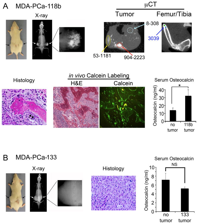
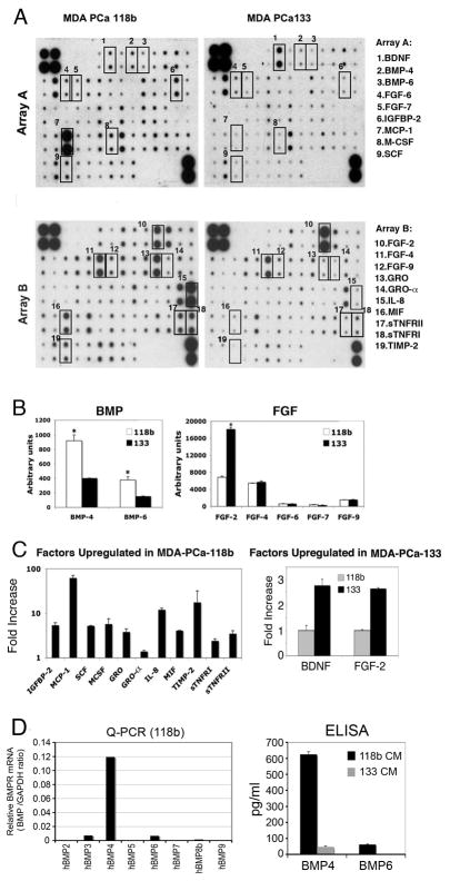
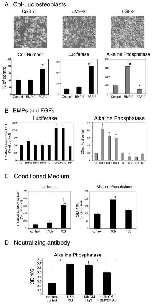
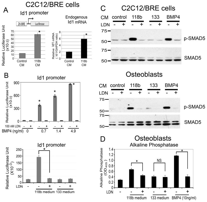
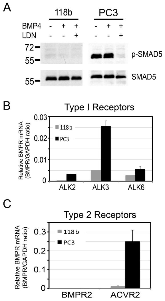
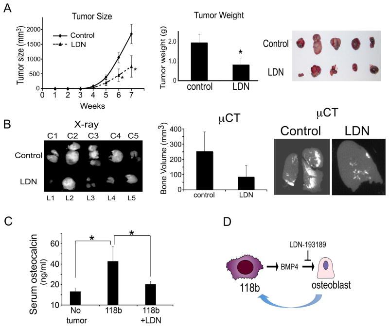
Induction of new bone formation is frequently seen in the bone lesions from prostate cancer. However, whether osteogenesis is necessary for prostate tumor growth in bone is unknown. Recently, 2 xenografts, MDA-PCa-118b and MDA-PCa-133, were generated from prostate cancer bone metastases. When implanted subcutaneously in severe combined immunodeficient (SCID) mice, MDA-PCa-118b induced strong ectopic bone formation while MDA-PCa-133 did not. To identify the factors that are involved in bone formation, we compared the expression of secreted factors (secretome) from MDA-PCa-118b and MDA-PCa-133 by cytokine array. We found that the osteogenic MDA-PCa-118b xenograft expressed higher levels of bone morphogenetic protein BMP4 and several cytokines including interleukin-8, growth-related protein (GRO), and CCL2. We showed that BMP4 secreted from MDA-PCa-118b contributed to about a third of the osteogenic differentiation seen in MDA-PCa-118b tumors. The conditioned media from MDA-PCa-118b induced a higher level of osteoblast differentiation, which was significantly reduced by treatment with BMP4 neutralizing antibody or the small molecule BMP receptor 1 inhibitor LDN-193189. BMP4 did not elicit an autocrine effect on MDA-PCa-118b, which expressed low to undetectable levels of BMP receptors. Treatment of SCID mice bearing MDA-PCa-118b tumors with LDN-193189 significantly reduced tumor growth. Thus, these studies support a role of BMP4-mediated osteogenesis in the progression of prostate cancer in bone.


Figures

Figure 1
Figure 1
Analysis of PCa-118b and PCa-133 xenografts. (A) Subcutaneously implanted PCa-118b tumor showed mineralization (arrows) within the tumor by X-ray and microCT analyses. Three varying bone densities within the tumor are shown in comparison with the uniformed, high bone density in the femur/tibia. Histological analysis showed the presence of mineralized bone and activated osteoblasts (arrows) around the bone. Calcein labeling demonstrated the irregularity of newly formed bone. Serum osteocalcin levels are significantly higher in PCa-118b tumor bearing mice compared to those of controls. (B) Subcutaneously implanted PCa-133 tumor (arrows) did not show mineralized bone radiographically or histologically. Serum osteocalcin levels in PCa-133 tumor bearing mice were not significantly (NS) different from those in control mice. * p < 0.05.
Figure 2
Figure 2
Cytokine expression profile of conditioned media from PCa-118b and PCa-133 xenografts. (A) Conditioned media from PCa-118b or PCa-133 were incubated with human antibody arrays that detected 120 cytokines. Cytokines that were differentially expressed are boxed. (B) The relative density of the array signals for BMPs and FGFs in the conditioned media of PCa-118b and PCa-133 xenografts was determined by Image J and expressed as arbitrary units. * p < 0.05. (C) Cytokines upregulated in PCa-118b versus PCa-133 conditioned media were determined by Image J and expressed as fold increase. Similar results were obtained using conditioned medium prepared from three different batches of xenograft tumors. (D) BMP RNA transcripts in PCa-118b or PCa-133 tumors were determined by qRT-PCR using primers specific to human BMPs (see primer list in Supplemental Table 1). Levels of human BMP4 and BMP6 proteins in PCa-118b and MDA-PCa-133 conditioned media were determined by ELISA.
Figure 3
Figure 3
BMPs, FGFs, and PCa-118b and PCa-133 conditioned media on osteoblast proliferation or differentiation. (A) Calvarial osteoblasts from Col-luc mice were incubated without or with BMPs (100 ng/ml) or FGF-2 (10 ng/ml) for three days. Cell morphology (x100), cell numbers, luciferase activity and alkaline phosphatase activity were measured. (B) BMP-2, BMP-4, BMP-6, FGF2, and FGF9 on Col-luc osteoblasts. (C) Conditioned media from PCa-118b or PCa-133 xenografts on Col-luc osteoblasts. (D) Neutralizing anti-BMP2/4 antibody on Col-luc osteoblasts. Each experiment was performed in triplicates and repeated at least three times. The averages of triplicates ± SD were shown. Data in A–C were expressed as a percent of control. * p < 0.05.
Figure 4
Figure 4
PCa-118b conditioned medium on BMP receptor signaling. (A) C2C12/BRE cells stably expressing a BMP-responsive Id1 promoter-luciferase reporter were incubated with PCa-118b conditioned medium for 12 hrs. Cell lysates were measured directly for luciferase activity. Endogenous Id1 and GAPDH messages were analyzed by qRT-PCR and expressed as Id1/GAPDH ratio. (B) C2C12/BRE cells were treated with increasing concentrations of either BMP4 (upper) or conditioned media from PCa-118b or PCa-133 (lower) in the presence or absence of 100 nM LDN-193189 for 12 hrs, and luciferase activities were analyzed. (C) C2C12/BRE cells (upper) or primary osteoblasts (lower) were treated as in B, and cell lysates were immunoblotted for phosphorylated Smad5 followed by reblotting for total Smad5. (D) Primary mouse calvarial osteoblasts were treated as in C except that alkaline phosphatase activity was determined after 3 days. * p < 0.05; NS, not significant.
Figure 5
Figure 5
Lack of BMP4 signaling on PCa-118b cells. (A) PCa-118b cells and PC3 were treated with BMP4 with or without LDN-193189 for 12 hrs, and analyzed for Smad5 phosphorylation. Levels of BMP Type I receptor (ALK2, 3, 6) (B) and Type II receptor (BMPR2, ACVRs) (C) transcripts in PCa-118b tumors and PC-3 cells were determined by qRT-PCR (see Supplemental Table 1).
Figure 6
Figure 6
LDN-193189 treatment attenuates PCa-118b tumor growth in vivo. (A) Cells were isolated from PCa-118b tumors, mixed 1:1 with matrigel, and injected into SCID mice subcutaneously (1 × 10⁶ cells per mouse). LDN-193189 or vehicle treatment was started five days post-tumor cell injection. Tumor sizes were measured weekly. At week 7, tumors were removed and weighed. (B) Tumors from vehicle and LDN-193189-treated groups were X-rayed. Bone volumes were determined by microCT using a 300 value as the cut-off. (C) Serum osteocalcin levels were determined for non-tumor bearing (n=5), PCa-118b tumor bearing (n=10), and PCa-118b tumor bearing but LDN-193189-treated (n=10) mice. (D) Model of bi-directional effect between PCa-118b osteogenic tumor and osteoblasts. PCa-118b secretes BMP4 that induces osteoblast differentiation. Inhibition of the paracrine BMP4 effect on osteoblasts leads to the attenuation of PCa-118b tumor growth, suggesting that osteoblasts play a role in supporting PCa-118b tumor growth.

References

    1. Jacobs SC. Spread of prostatic cancer to bone. Urology. 1983;21:337–44. - PubMed
    1. Shah RB, Mehra R, Chinnaiyan AM, Shen R, Ghosh D, Zhou M, et al. Androgen-independent prostate cancer is a heterogeneous group of diseases: lessons from a rapid autopsy program. Cancer Res. 2004;64:9209–16. - PubMed
    1. Saad F, Lipton A. Bone-marker levels in patients with prostate cancer: potential correlations with outcomes. Curr Opin Support Palliat Care. 2010;4:127–34. - PubMed
    1. Logothetis C, Lin S-H. Osteoblasts in prostate cancer metastasis to bone. Nature Reviews Cancer. 2005;5:21–8. - PubMed
    1. Li Z, Mathew P, Yang J, Starbuck M-W, Zurita AJ, Liu J, et al. Androgen receptor–negative human prostate cancer cells induce osteogenesis through FGF9-mediated mechanisms. J Clin Invest. 2008;118:2697–710. - PMC - PubMed

Publication types

MeSH terms