. 2011 Jul 1;124(Pt 13):2208-19.
doi: 10.1242/jcs.080523.

Proteolytically cleaved MLL subunits are susceptible to distinct degradation pathways

Affiliations

Proteolytically cleaved MLL subunits are susceptible to distinct degradation pathways

Akihiko Yokoyama et al. J Cell Sci. .

Abstract

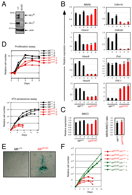
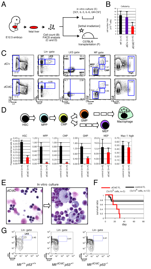
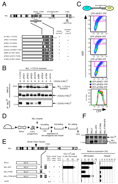
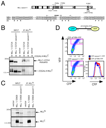
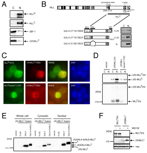
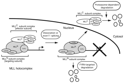
The mixed lineage leukemia (MLL) proto-oncogenic protein is a histone-lysine N-methyltransferase that is produced by proteolytic cleavage and self-association of the respective functionally distinct subunits (MLL(N) and MLL(C)) to form a holocomplex involved in epigenetic transcriptional regulation. On the basis of studies in Drosophila it has been suggested that the separated subunits might also have distinct functions. In this study, we used a genetically engineered mouse line that lacked MLL(C) to show that the MLL(N)-MLL(C) holocomplex is responsible for MLL functions in various developmental processes. The stability of MLL(N) is dependent on its intramolecular interaction with MLL(C), which is mediated through the first and fourth plant homeodomain (PHD) fingers (PHD1 and PHD4) and the phenylalanine/tyrosine-rich (FYRN) domain of MLL(N). Free MLL(N) is destroyed by a mechanism that targets the FYRN domain, whereas free MLL(C) is exported to the cytoplasm and degraded by the proteasome. PHD1 is encoded by an alternatively spliced exon that is occasionally deleted in T-cell leukemia, and its absence produces an MLL mutant protein that is deficient for holocomplex formation. Therefore, this should be a loss-of-function mutant allele, suggesting that the known tumor suppression role of MLL may also apply to the T-cell lineage. Our data demonstrate that the dissociated MLL subunits are subjected to distinct degradation pathways and thus not likely to have separate functions unless the degradation mechanisms are inhibited.


Figures

Fig. 1.
Fig. 1.
MLLC is required for MLL-dependent transcription during embryogenesis. (A) Schematic representations of dC-mutant MLL proteins. (B) The structure of the targeting vector. The positions of diagnostic primers are shown at the bottom. (C) Successful recombination of positive ES clones was confirmed by PCR followed by digestion with XhoI. The XhoI site downstream of exon 28 was destroyed in the recombined allele as shown in B. (D) Sequences of PCR fragments amplified from genomic DNAs of the recombined ES clones. (E) Expression of MLL proteins in embryos at E11.5. Whole-embryo extracts were immunoblotted with anti-MLLN (mmN4.4), anti-MLLC (9–12) or anti-menin antibody. The diagnostic PCR data are shown in the bottom panel. (F) Genotypes at various developmental stages. The numbers of embryos or mice with the indicated genotypes are shown for each developmental stage. Viability was confirmed by presence of a beating heart. (G) Abnormal features of dC/dC embryo at E12.5. (H) Expression of Hoxc8 transcripts in E10.5 embryos. Whole-mount in situ hybridization was performed using antisense Hoxc8 probes (Hoxc8 AS). Arrows indicate positions of target gene expression.
Fig. 2.
Fig. 2.
The MLL holocomplex is required for MLL-dependent transcription in MEFs. (A) Protein expression of the MLL subunits in MEFs. Cell lysates prepared from wt or dC/dC MEFs were immunoblotted with anti-MLLN (mmN4.4), anti-MLLC(9–12) or anti-actin antibody. (B) Expression of various genes in dC/dC MEFs. Three independently established lines for the indicated genotypes were examined by quantitative RT-PCR for the genes indicated at the top of each panel. RNA was prepared from the third passage MEFs before they went into senescence. Relative expression levels (normalized to Gapdh) of various transcripts are depicted relative to those of clone #1, which were arbitrarily set as 1. Error bars represent the standard deviations of triplicate PCRs. (C) Expression of Mll was analyzed using a qPCR probe for a coding sequence downstream of the processing site as in B. The ratios of the Mll mRNA signal detected by the probe upstream of the processing site (N) toward that by the probe downstream of the processing site (C) are shown in the right panel. (D) Proliferation and 3T3 senescence assays were performed on wt and dC/dC MEFs. MEFs were analyzed after the third passage. A representative result, in which three clones each for the two genotypes were analyzed in a single experiment, is shown. Different clones were also analyzed and reproducibly showed a similar result. (E) Senescence-associated β-galactosidase assay of wt or dC/dC MEFs. (F) Proliferation and 3T3 senescence assays were performed for three lines each of dC/dC MEFs with or without homozygous p53 knockout alelles. MEFs were analyzed after the third passage.
Fig. 3.
Fig. 3.
The MLL holocomplex is required for HSC maintenance and expansion of hematopoietic progenitors, but not for differentiation. (A) The experimental scheme of B–F. (B) Cellularities of dC mutant fetal livers. Error bars represent the standard deviation of cell numbers of 4 or 6 livers. (C) Representative FACS profiles of E12.5 fetal liver cells. Lineage cocktail (anti-CD3, -CD4, -CD8, -B220, -TER119 and -Gr-1) was used to define lineage negative fractions. A marked decrease in the LKS and multipotent progenitor populations was observed in dC/dC livers. HSCs are defined as CD48 within the LKS gate where MPPs are defined as CD48+ in this study (Christensen and Weissman, 2001; Kim et al., 2006). It should, however, be noted that an alternative model has also been proposed (Mansson et al., 2007). (D) Average frequencies of various hematopoietic cell sub-populations per total liver cells. Controls include wt and dC/+. The number of embryos analyzed is indicated below. (E) The morphology of dC/dC fetal liver cells. Enucleated red blood cells were present in dC/dC fetal livers. Functional macrophages with engulfed materials emerged after 1 week in culture in methylcellulose medium containing GM-CSF, SCF, IL-3 and IL-6. (F) The ability of dC mutant fetal liver cells to reconstitute the hematopoietic system. Fetal liver cells (5×10⁵) from dC/dC embryos (n=5) or control embryos (5×10⁴; n=12) were injected into lethally irradiated recipients. The survival ratio during the monitoring period (80 days) is shown. (G) Hematopoietic defects of dC mutants are not caused by p53-dependent senescence. FACS plots of the fetal liver cells, with the various genotypes indicated below, are shown using the lineage cocktail, cKit and Sca1 antibodies.
Fig. 4.
Fig. 4.
PHD1 and PHD4 are required for intramolecular interaction in addition to FYRN, which also serves as a destabilization signal. (A) Schematic presentation of the various MLL 1/2254 substitution and deletion mutants analyzed. The binding property with MLLC and susceptibility to destabilization are shown on the right. (B) Immunoprecipitation (IP) and western blotting analysis was performed for various MLL 1/2254 mutants that express residues 1–2254 with the indicated substitution and deletion mutations. MLL 1/2254 mutants were coexpressed with Xpress-tagged GAL4-MLLC [(X)GAL4-MLLC] in 293T cells. The cell extracts were subjected to IP with anti-MLLN (mmN4.4) antibody followed by immunoblotting. The precipitates and input samples indicated on the left were immunoblotted with anti-MLLN (mmN4.4; top panel) and/or anti-Xpress (middle and bottom panels) antibodies. (C) Various CFP–MLL 34/3606–YFP mutants were transiently expressed in 293T cells and analyzed by flow cytometry. A population that highly expressed YFP was gated and shown in overlay histograms for its CFP expression level. (D) The experimental scheme for the myeloid progenitor transformation assay. Expression of Hoxa9 was analyzed at the end of the first round of plating. Colony forming units (CFUs) were measured at the end of the third round plating. (E) Schematic representation of the MLL–AF9 mutants with or without an FYRN domain. The destabilization property, Hoxa9 expression, and transformation ability are summarized. CFUs per 104 cells at the third round are shown, with error bars representing the standard deviations from three independent analyses. Relative expression levels (normalized to the β-actin gene) of Mll mutant and Hoxa9 transcripts in the first round colonies are depicted relative to MLL–AF9-transduced cells arbitrarily set as 100 (%). Quantitative PCR was performed with specific primers and probes for human MLL (which detects various MLL mutants but not endogenous mouse Mll) or mouse Hoxa9 and standardized to the β-actin gene. (F) Protein expression of various FYRN mutants. MLL–AF9 mutants fused with or without an FYRN domain were expressed in plat-E cells and immunoblotted with anti-MLLN antibody (mmN4.4).
Fig. 5.
Fig. 5.
The MLL Δexon11 mutant is incapable of forming an intramolecular MLL holocomplex and is susceptible to degradation. (A) Schematic representation of the Δexon11 mutant. The exon and intron structures around exon 11 are conserved between human (hMLL) and mouse (mMLL). (B) IP analysis was performed for the MLL 1/2254 Δexon11 mutant as in Fig. 4B. (C) IP analysis was performed for the MLL full-length Δexon11 mutant. IP analysis was performed for various MLL full-length Δexon11 tagged with an HA epitope at the C-terminus. MLL mutants were transiently expressed in 293T cells. The cell extracts were subjected to IP with anti-MLLN (mmN4) antibody followed by immunoblotting. The precipitates and input samples, indicated on the top, were immunoblotted with anti-MLLN (mmN4; top panel) and anti-HA(3F10) antibodies (bottom panel). (D) The stability of MLLN Δexon11 was analyzed as in Fig. 4C.
Fig. 6.
Fig. 6.
MLLC is exported to the cytosol after dissociation from MLLN. (A) Subcellular localization of the endogenous MLL proteins and exogenous MLLC. Xpress–FLAG-tagged (Xf) MLLC (or vector) was transiently expressed in 293T cells that were lysed and separated into cytosolic (C) and nuclear (N) fractions and then immunoblotted for the indicated proteins with anti-MLLN (mmN4.4), anti-MLLC (mmC2.1), anti-SBF-1 (vector-transfected cells: upper three panels) or anti-Xpress antibodies [(Xf)MLLC construct-transfected cells: bottom panel]. Sbf-1 served as a control for cytosolic localization. (B) Subcellular localization of various GAL4-MLLC mutants. Various GAL4–MLL mutants, which are schematically illustrated, were analyzed for their subcellular localization as in A. GAL4 fusion proteins were visualized by anti-GAL4 antibody. (C) Colocalization of exogenous MLLN or MLLN Δexon11 with MLLC. The indicated MLLN mutants and (Xf)MLLC fragments were coexpressed in 293T cells and analyzed by indirect immunofluorescence with anti-MLLN (rpN1) and anti-Xpress antibodies. The MLLN Δexon11 mutant served as a negative control. (D) Subcellular localization of exogenous full-length MLL proteins with or without the Δexon11 mutation. Full-length MLL tagged with Xpress and FLAG at its N-terminus and HA at its C-terminus [(Xf)MLL(H)] with or without deletion of exon 11 was analyzed as in A. Each fraction was immunoblotted with anti-Xpress or anti-HA antibody. (E) Expression of various MLLC mutants driven by the same promoter and translation initiation sites with or without additional nuclear localization signals. (Xf)MLLC, (X)GAL4–MLLC, (X)GAL4–x3NLS–MLLC were expressed in 293T cells, fractionated and immunoblotted with anti-Xpress antibody. (F) Sensitivities of MLLN and MLLC to the MG132 proteasome inhibitor. 293T cells were transfected with the corresponding expression vectors and cultured with and without 10 μM MG132 for 8 hours and subjected to western blotting with anti-Xpress antibody. A nonspecific band (nsp) serves as a loading control.
Fig. 7.
Fig. 7.
A model of the two distinct degradation pathways for each MLL subunit.

References

    1. Alvarez-Venegas R., Avramova Z. (2001). Two Arabidopsis homologs of the animal trithorax genes: a new structural domain is a signature feature of the trithorax gene family. Gene 271, 215-221 - PubMed
    1. Ayton P. M., Cleary M. L. (2001). Molecular mechanisms of leukemogenesis mediated by MLL fusion proteins. Oncogene 20, 5695-5707 - PubMed
    1. Ayton P. M., Chen E. H., Cleary M. L. (2004). Binding to nonmethylated CpG DNA is essential for target recognition, transactivation, and myeloid transformation by an MLL oncoprotein. Mol. Cell. Biol. 24, 10470-10478 - PMC - PubMed
    1. Bertolino P., Tong W. M., Galendo D., Wang Z. Q., Zhang C. X. (2003). Heterozygous Men1 mutant mice develop a range of endocrine tumors mimicking multiple endocrine neoplasia type 1. Mol. Endocrinol. 17, 1880-1892 - PubMed
    1. Birke M., Schreiner S., Garcia-Cuellar M. P., Mahr K., Titgemeyer F., Slany R. K. (2002). The MT domain of the proto-oncoprotein MLL binds to CpG-containing DNA and discriminates against methylation. Nucleic Acids Res. 30, 958-965 - PMC - PubMed

Publication types

MeSH terms