. 2011 Jun 13;193(6):1033-48.
doi: 10.1083/jcb.201101056.

The cortical protein Lte1 promotes mitotic exit by inhibiting the spindle position checkpoint kinase Kin4

Affiliations

The cortical protein Lte1 promotes mitotic exit by inhibiting the spindle position checkpoint kinase Kin4

Daniela Trinca Bertazzi et al. J Cell Biol. .

Abstract

The spindle position checkpoint (SPOC) is an essential surveillance mechanism that allows mitotic exit only when the spindle is correctly oriented along the cell axis. Key SPOC components are the kinase Kin4 and the Bub2-Bfa1 GAP complex that inhibit the mitotic exit-promoting GTPase Tem1. During an unperturbed cell cycle, Kin4 associates with the mother spindle pole body (mSPB), whereas Bub2-Bfa1 is at the daughter SPB (dSPB). When the spindle is mispositioned, Bub2-Bfa1 and Kin4 bind to both SPBs, which enables Kin4 to phosphorylate Bfa1 and thereby block mitotic exit. Here, we show that the daughter cell protein Lte1 physically interacts with Kin4 and inhibits Kin4 kinase activity. Specifically, Lte1 binds to catalytically active Kin4 and promotes Kin4 hyperphosphorylation, which restricts Kin4 binding to the mSPB. This Lte1-mediated exclusion of Kin4 from the dSPB is essential for proper mitotic exit of cells with a correctly aligned spindle. Therefore, Lte1 promotes mitotic exit by inhibiting Kin4 activity at the dSPB.


Figures

Figure 1.
Figure 1.
Lte1 interacts with Kin4 in vitro and in vivo. (A) Kin4- and Lte1-interacting partners found by MS analysis. (B–E) Kin4 interacts with Lte1 and Kel1. Immunoprecipitations using anti-HA or anti-Myc beads as indicated. (F and G) In vitro binding assay using bacterially expressed KIN4 and LTE1. (F) Lte1 truncations used in G; numbers represent amino acid positions. (G) Immunoblotting of bacterially expressed GST (lane 1) and GST-Kin4 (lane 2) bound to glutathione-Sepharose beads. Protein extracts of E. coli expressing 6His (lane 3), 6His-Lte1-N (lane 4), and 6His-Lte1-C (lane 5) were incubated with GST (lanes 6, 8, and 10) or GST-Kin4 (lanes 7, 9, and 11) for 2 h at 4°C.
Figure 2.
Figure 2.
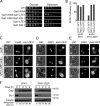
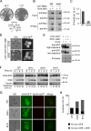
Lte1 inhibits Kin4 function toward Bfa1 in vivo. (A) Co-overexpression of LTE1 rescues the lethality arising from Gal1-KIN4 overexpression. Serial dilutions of cells spotted on YPAR plates containing either glucose or galactose to inhibit or induce the expression of Gal1 promoter, respectively. (B) SPOC proficiency in the indicated strains grown at 23°C and shifted to 30°C for 4 h before inspection. One representative experiment of three is shown. (C–E) kar9Δ or SFK1-GBP kar9Δ cells expressing LTE1-GFP (C), KEL1-GFP (D), and KEL2-GFP (E) were analyzed by fluorescence microscopy after incubation for 4 h at 30°C. Mother (M) and daughter (D) cell bodies are indicated. Cells in anaphase with correctly aligned or misoriented spindles are shown. The cell with two buds in SFK1-GBP kar9Δ LTE1-GFP is indicative of inappropriate mitotic exit. DNA was DAPI stained. Bars, 5 µm. (F) SFK1 and SFK1-GBP cells carrying LTE1-GFP were arrested with α-factor and released into nocodazole-containing media. Clb2 and Sic1 levels were determined by immunoblotting at the indicated time points. Tubulin served as a loading control.
Figure 3.
Figure 3.
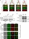
Kin4 catalytic kinase activity is required for Kin4–Lte1 interaction. (A) Representative frames and fluorescence intensity line traces of cells expressing KIN4-GFP LTE1-3Cherry. Line profiles above the images represent the fluorescence intensities (FI) in arbitrary units (A.U.), measured for the indicated rectangular area, for Kin4-GFP (green lines) and Lte1-3Cherry (red lines). Note that FI is not comparable between cells. Cell boundaries in each graph are indicated as M and D, and bud neck region as BN. Bar, 5 µm. (B) LTE1 and LTE1-9Myc strains carrying KIN4-6HA (WT), KIN4-6HA elm1Δ, and KIN4-T209A-6HA were subjected to immunoprecipitation using anti-Myc beads. (C) Interaction between Lte1-9Myc and Kin4-6HA was investigated in RTS1 (WT, wild type) and rts1Δ cells upon immunoprecipitation of Lte1-9Myc with anti-Myc beads. (D) Localization of Kin4-GFP and Kin4-T209A-GFP in strains carrying LTE1-3Cherry (no overexpression) and Gal1-LTE1-3Cherry (overexpression), growing in galactose-containing medium. Bars, 5 µm.
Figure 4.
Figure 4.
Lte1 inhibits the catalytic activity of Kin4 in vitro. (A) Yeast-purified GST-Kin4 or GST-Kin4-T209A were incubated with kinase buffer (lane 1) or purified 6His-GFP (lane 2) in the absence or presence of increasing amounts of 6His-Lte1-N (B) or 6His-Lte1-C (C) as indicated. Reactions were preincubated for 5 min at 30°C before adding MBP-Bfa1 and further incubating for 30 min at the same temperature. Autoradiographs (32P), Coomassie-stained protein gels, and specific Kin4 kinase activity (in arbitrary units) are shown. Molar ratios were calculated based on protein molecular weight and concentration used in each reaction. One out of two experiments is shown.
Figure 5.
Figure 5.
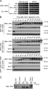
Functional relationship between Kin4 and Lte1 during normal cell cycle progression. (A) lte1Δ cells become more sensitive to increasing KIN4 levels. The indicated strains carrying LTE1 on a URA3-based plasmid were transformed with KIN4 on a LEU2-based centromeric (CEN-KIN4) or 2µ-based plasmid (2µ-KIN4). Shown is the growth (2 d at 30°C) of serial dilutions of cells. (B) The indicated strains were arrested in G1 with α-factor in galactose-containing medium at 30°C. Cells were washed with glucose-containing medium to remove α-factor (t = 0) and to simultaneously induce UPL-Tem1 degradation. Bfa1-3HA, Clb2, and Upl-Tem1 were detected using anti-HA, anti-Clb2, and anti-Tem1 antibodies, respectively. Asterisks indicate Bfa1-3HA hyperphosphorylation forms. “C” is a control sample containing hyperphosphorylated Bfa1-3HA (enriched from cdc15-1 cells arrested in late anaphase; Pereira and Schiebel, 2005). Tubulin served as loading control. (C) Indicated strains carrying BFA1-3HA were arrested with α-factor and released in α-factor–free medium. Samples of the indicated time points were run side by side for comparison of Bfa1 phosphorylation (see Fig. S4, C–-F, for complete samples). The asterisk indicates Cdc5-dependent hyperphosphorylated Bfa1 form.
Figure 6.
Figure 6.
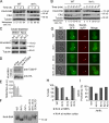
Lte1 regulates the phosphorylation status of Kin4. (A and B) Phosphorylation of Kin4-6HA in the presence or absence of LTE1. (A) Cells carrying KIN4-6HA were synchronized in G1 with α-factor (t = 0) and released into media containing nocodazole to induce metaphase arrest and Kin4 phosphorylation (asterisk). (B) Nocodazole-arrested cells were released in nocodazole-free medium. Clb2 levels and percentage of metaphase cells were monitored over time. (C) Kin4-6HA of wild-type and lte1Δ cells arrested in metaphase upon CDC20 depletion in the presence of solvent control (DMSO) or nocodazole. Asterisks mark Kin4-phosphorylated forms. Tubulin in A–C served as loading control. (D) KIN4-6HA, KIN4-6HA lte1Δ, and kin4-T209A-6HA strains, arrested in metaphase (CDC20 depletion), were subjected to immunoprecipitation using anti-HA antibodies. The levels of Kin4-6HA and phosphorylation at T209 are shown. (E) Quantification of D. (F) Strains expressing KIN4-6HA were arrested in G1 with α-factor and released into medium containing nocodazole for 2 h at 30°C. CEN-LTE1 indicates a centromeric plasmid carrying LTE1. Asterisk points to the Kin4-6HA–hyperphosphorylated form. (G) Kin4-GFP localization was determined by fluorescence microscopy in nocodazole-treated metaphase-arrested cells. Spc42-eqFP served as SPB marker (arrowheads). Bar, 5 µm. (H) Quantification of G. (I) SPOC proficiency of the indicated strains grown at 23°C and shifted to 30°C for 4 h before inspection. G and I show one representative experiment of three. 100–150 cells were scored per sample.
Figure 7.
Figure 7.
Lte1 regulates loading of Kin4 onto the dSPB. (A) Localization of Kin4-GFP monitored by fluorescence microscopy using unfixed cells. Spc42-eqFP served as SPB marker (arrowhead). Note that the SPB retained in the mother cell (new SPB) is weakly labeled due to the slow maturation property of Spc42-eqFP (Pereira et al., 2001). Bar, 5 µm. (B) Quantification of A; 100–150 cells were scored per strain. One representative experiment of three is shown.
Figure 8.
Figure 8.
Lte1–Kin4 interaction requires Cla4. (A) Growth of the indicated strains in YPD plates for 2 d (30°C) and 14 d (11°C). (B) Interaction between Lte1-9Myc and Kin4-6HA was investigated in wild-type (WT) and cla4Δ cells upon immunoprecipitation of Lte1-9Myc with anti-Myc beads. Note that this experiment was done with the experiment shown in Fig. 3 C; i.e., the blots of the WT strain are identical. (C) Quantification of B. One representative experiment of two is shown. (D) Localization of Lte1-GFP. (E) Phosphorylation profile of Lte1-GFP in the indicated strains. The brackets indicate Lte1-phosphorylated forms. (F) Strains were arrested with α-factor and released into nocodazole-containing media. Kin4-6HA and Clb2 levels were determined by immunoblotting at the indicated time points. Tubulin served as a loading control. (G) Localization of Kin4-GFP. Spc42-eqFP served as SPB marker. (H) Quantification of G. One representative experiment of three is shown. Bar, 5 µm.
Figure 9.
Figure 9.
Model for Lte1 regulation of mitotic exit. (A) Lte1 functions upstream of Tem1 to promote mitotic exit. Both Elm1 and Rts1 control Kin4 activity by promoting Kin4 catalytic activity and localization, respectively. In contrast, Lte1 inhibits Kin4 catalytic activity, working upstream of Tem1. The kinase Cla4 is required for Lte1 to interact with Kin4. (B) Spatial regulation of Kin4 by Lte1. Cla4 activates Lte1 to inhibit the binding of Kin4 at dSPB to facilitate mitotic exit. See Discussion for details.

Similar articles

Cited by

References

    1. Adames N.R., Oberle J.R., Cooper J.A. 2001. The surveillance mechanism of the spindle position checkpoint in yeast. J. Cell Biol. 153:159–168 10.1083/jcb.153.1.159 - DOI - PMC - PubMed
    1. Audhya A., Emr S.D. 2002. Stt4 PI 4-kinase localizes to the plasma membrane and functions in the Pkc1-mediated MAP kinase cascade. Dev. Cell. 2:593–605 10.1016/S1534-5807(02)00168-5 - DOI - PubMed
    1. Bardin A.J., Amon A. 2001. Men and sin: what’s the difference? Nat. Rev. Mol. Cell Biol. 2:815–826 10.1038/35099020 - DOI - PubMed
    1. Bardin A.J., Visintin R., Amon A. 2000. A mechanism for coupling exit from mitosis to partitioning of the nucleus. Cell. 102:21–31 10.1016/S0092-8674(00)00007-6 - DOI - PubMed
    1. Carminati J.L., Stearns T. 1997. Microtubules orient the mitotic spindle in yeast through dynein-dependent interactions with the cell cortex. J. Cell Biol. 138:629–641 10.1083/jcb.138.3.629 - DOI - PMC - PubMed

Publication types

MeSH terms

Substances