. 2011 Jun 28;108(26):10484-9.
doi: 10.1073/pnas.1017029108. Epub 2011 Jun 13.

Structural and functional characterization of an atypical activation domain in erythroid Kruppel-like factor (EKLF)

Affiliations

Structural and functional characterization of an atypical activation domain in erythroid Kruppel-like factor (EKLF)

Caroline Mas et al. Proc Natl Acad Sci U S A. .

Abstract

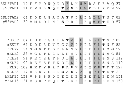
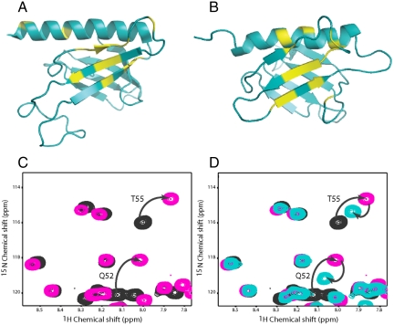
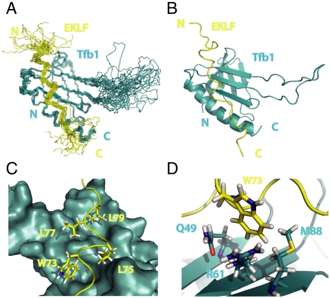
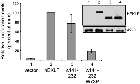
Erythroid Krüppel-like factor (EKLF) plays an important role in erythroid development by stimulating β-globin gene expression. We have examined the details by which the minimal transactivation domain (TAD) of EKLF (EKLFTAD) interacts with several transcriptional regulatory factors. We report that EKLFTAD displays homology to the p53TAD and, like the p53TAD, can be divided into two functional subdomains (EKLFTAD1 and EKLFTAD2). Based on sequence analysis, we found that EKLFTAD2 is conserved in KLF2, KLF4, KLF5, and KLF15. In addition, we demonstrate that EKLFTAD2 binds the amino-terminal PH domain of the Tfb1/p62 subunit of TFIIH (Tfb1PH/p62PH) and four domains of CREB-binding protein/p300. The solution structure of the EKLFTAD2/Tfb1PH complex indicates that EKLFTAD2 binds Tfb1PH in an extended conformation, which is in contrast to the α-helical conformation seen for p53TAD2 in complex with Tfb1PH. These studies provide detailed mechanistic information into EKLFTAD functions as well as insights into potential interactions of the TADs of other KLF proteins. In addition, they suggest that not only have acidic TADs evolved so that they bind using different conformations on a common target, but that transitioning from a disordered to a more ordered state is not a requirement for their ability to bind multiple partners.


Conflict of interest statement

The authors declare no conflict of interest.

Figures

Fig. 1.
Fig. 1.
EKLFTAD1 and EKLFTAD2 are homologous to p53TAD1 and p53TAD2. (Top) Sequence alignments of EKLFTAD1 and EKLFTAD2 from human EKLF with p53TAD1 and p53TAD2 from human p53. The residues of p53 that form helices in the p53TAD1/MDM2 complex (23) and the p53TAD2/Tfb1PH complex (27) are underlined in black. The hydrophobic residues at the binding interfaces of the p53TAD1/MDM2 complex and the p53TAD2/Tfb1PH are in gray. Several known or potential phosphorylation sites are in bold. (Lower) Sequence alignment of regions of other KLF proteins (mouse and human) that share homology with EKLFTAD2. The KLF proteins are members of either KLF group 3 (EKLF, KLF2, KLF4) or KLF group 4 (KLF5, KLF15) based on phylogenetic analysis (1). Key hydrophobic residues are shaded gray and potential phosphorylation sites are in bold.
Fig. 2.
Fig. 2.
EKLFTAD2 and p53TAD2 share a common binding site on Tfb1PH/p62PH. Ribbon models of the 3D structures of Tfb1PH (A) and p62PH (B). The amino acids showing a significant chemical shift change (Δδ(ppm) > 0.15; Δδ = [(0.17ΔNH)2 + (ΔHN)2]1/2) upon formation of a complex with EKLFTAD2 are shown in yellow (A and B). (C) Overlay of a selected region from the two-dimensional 1H-15N HSQC spectra for a 0.5-mM sample of 15N-labeled p53TAD2 in the free form (black) and in the presence of 0.4 mM unlabeled Tfb1PH (pink). (D) Overlay of a selected region from the two-dimensional 1H-15N HSQC spectra for a 0.5-mM sample of 15N-labeled p53TAD2 in the free form (black), in the presence of 0.4 mM unlabeled Tfb1PH (pink), and after addition of 0.5 mM unlabeled EKLFTAD2 (aqua). Signals that undergo significant changes in 1H and 15N chemical shifts upon formation of the complex with Tfb1PH (C) and that return toward their original position following the addition of EKLFTAD2 (D) are indicated by arrows.
Fig. 3.
Fig. 3.
Structure of the Tfb1PH/EKLFTAD2 Complex. (A) Overlay of the 20 lowest-energy structures of the complex between Tfb1PH (blue) and EKLFTAD2 (yellow). The structures were superimposed using the backbone atoms C′, Cα, and N of residues 4–65 and 85–112 of Tfb1PH and residues 59–84 of EKLFTAD2. (B) Ribbon model of the lowest-energy conformer of the complex between Tfb1PH (blue) and EKLFTAD2 (yellow). (C) Three-dimensional structure of Tfb1PH is shown as molecular surface (blue), and EKLFTAD2 is represented as a tube (yellow). The side chains of several hydrophobic residues of EKLFTAD2 (Trp73, Leu75, Leu77, and Leu79) that form the interface with Tfb1PH are shown as sticks. (D) Three-dimensional structure of Tfb1PH is shown as ribbon (blue), and EKLFTAD2 is represented as a tube (yellow). The side chain of Trp73 (EKLFTAD2) is positioned in a pocket formed by Gln49, Arg61, and M88 of Tfb1.
Fig. 4.
Fig. 4.
Trp73 influences in vivo activation of β-globin gene expression. K562 cells were cotransfected with plasmids expressing the luciferase reporter gene under control of ß-globin promoter along with the EKLF constructs (EKLF, EKLFΔ140–232, and EKLFW73PΔ140–232). A plasmid expressing Renilla luciferase was included as a control for normalization of transfection efficiency. Transcriptional activities were normalized to that of full-length hEKLF, which was set at 100%. Error bars represent standard error of the mean of three independent experiments, each performed in triplicate. (Inset) The level of expression of the proteins after transfection into Cos7 cells as monitored by Western blot with an antibody against EKLF. Actin levels are shown as a loading control.

References

    1. Bieker JJ. Kruppel-like factors: Three fingers in many pies. J Biol Chem. 2001;276:34355–34358. - PubMed
    1. Kaczynski J, Cook T, Urrutia R. Sp1- and Kruppel-like transcription factors. Genome Biol. 2003;4(2):206. - PMC - PubMed
    1. Miller IJ, Bieker JJ. A novel, erythroid cell-specific murine transcription factor that binds the CACCC element and is related to the Kruppel family of nuclear proteins. Mol Cell Biol. 1993;13:2776–2786. - PMC - PubMed
    1. Hodge D, et al. A global role for EKLF in definitive and primitive erythropoiesis. Blood. 2006;107:3359–3370. - PMC - PubMed
    1. Perkins AC, Sharpe AH, Orkin SH. Lethal beta-thalassaemia in mice lacking the erythroid CACCC-transcription factor EKLF. Nature. 1995;375:318–322. - PubMed

Publication types

Substances

Associated data