. 2011 Jun 28;108(26):10696-701.
doi: 10.1073/pnas.1104306108. Epub 2011 Jun 13.

Crystal structure of mouse coronavirus receptor-binding domain complexed with its murine receptor

Affiliations

Crystal structure of mouse coronavirus receptor-binding domain complexed with its murine receptor

Guiqing Peng et al. Proc Natl Acad Sci U S A. .

Abstract

Coronaviruses have evolved diverse mechanisms to recognize different receptors for their cross-species transmission and host-range expansion. Mouse hepatitis coronavirus (MHV) uses the N-terminal domain (NTD) of its spike protein as its receptor-binding domain. Here we present the crystal structure of MHV NTD complexed with its receptor murine carcinoembryonic antigen-related cell adhesion molecule 1a (mCEACAM1a). Unexpectedly, MHV NTD contains a core structure that has the same β-sandwich fold as human galectins (S-lectins) and additional structural motifs that bind to the N-terminal Ig-like domain of mCEACAM1a. Despite its galectin fold, MHV NTD does not bind sugars, but instead binds mCEACAM1a through exclusive protein-protein interactions. Critical contacts at the interface have been confirmed by mutagenesis, providing a structural basis for viral and host specificities of coronavirus/CEACAM1 interactions. Sugar-binding assays reveal that galectin-like NTDs of some coronaviruses such as human coronavirus OC43 and bovine coronavirus bind sugars. Structural analysis and mutagenesis localize the sugar-binding site in coronavirus NTDs to be above the β-sandwich core. We propose that coronavirus NTDs originated from a host galectin and retained sugar-binding functions in some contemporary coronaviruses, but evolved new structural features in MHV for mCEACAM1a binding.


Conflict of interest statement

The authors declare no conflict of interest.

Figures

Fig. 1.
Fig. 1.
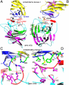
Structure of MHV-NTD/mCEACAM1a complex. (A) Domain structure of MHV spike protein. NTD: N-terminal domain; RBD: receptor-binding domain; HR-N: heptad-repeat N; HR-C: heptad-repeat C; TM: transmembrane anchor; IC: intracellular tail. The signal peptide corresponds to residues 1–14 and is cleaved during molecular maturation (45). Structures and functions of gray areas have not been clearly defined. (B) Kinetics and binding affinity of NTD and mCEACAM1a. (C) Structure of NTD/mCEACAM1a complex. Two β-sheets of the NTD core are in green and magenta, respectively; receptor-binding motifs (RBMs) are in red; other parts of the NTD are in cyan; mCEACAM1a is in yellow; and virus-binding motifs (VBMs) are in blue. N*: N terminus; C*: C terminus. (D) Sequence and secondary structures of NTD. β-Strands are shown as arrows, and the disordered region as a dashed line.
Fig. 2.
Fig. 2.
Structural details of MHV-NTD/mCEACAM1a interface. (A) Another view of the MHV-NTD/mCEACAM1a structure, which is derived by rotating the one in Fig. 1C 90° clockwise along a vertical axis. Virus-binding motif 1 (VBM1) on MHV NTD includes strands βC, βC′, and βC′′ and loops CC′, C′C′′, and C′′D. VBM2 on MHV NTD corresponds to loop FG. (B) Distribution of glycosylation sites and disulfide bonds. Glycans and glycosylated asparagines are in magenta, and cysteines are in yellow. The orientation of the structure is the same as in Fig. 1C. (C) A hydrophobic patch at the interface that is important for MHV-NTD/mCEACAM1a binding. MHV residues are in magenta, and mCEACAM1a residues are in green. The orientation of the structure is derived by rotating the structure in Fig. 1C 180° along a vertical axis. (D) Another hydrophobic patch at the interface that is important for MHV-NTD/mCEACAM1a binding. The orientation of the structure is slightly adjusted from the one in Fig. 1C.
Fig. 3.
Fig. 3.
Sequence analysis and mutagenesis studies of coronavirus/CEACAM1 interactions. (A) List of contact residues at the interface. (B) Partial sequence alignment of group 2a coronavirus NTDs. Contact residues are in red, important noncontact residues are in blue, and loop 10–11 is in green. Asterisks indicate positions that have fully conserved residues. Colons indicate positions that have strongly conserved residues. Periods indicate positions that have weakly conserved residues. (C) Partial sequence alignment of mammalian CEACAM1 proteins. (D) Structure-guided mutagenesis data on MHV NTD. Measured was mCEACAM1a-dependent cell entry by lentiviruses pseudotyped with wild-type or mutant MHV-A59 spike proteins. SEs are shown. (E) Published mutagenesis data on MHV NTD (28, 36, 37).
Fig. 4.
Fig. 4.
Structural comparisons of MHV NTD, human galectins, and rotavirus VP4. (A) MHV NTD. The orientation of the structure is the same as in Fig. 1C. (B) Human galectin-3 [Protein Data Bank (PDB) 1A3K]. The β-sandwich core is labeled and colored the same as in MHV NTD. Bound galactose is in yellow. (C) Rotavirus VP4 (PDB 1KQR). Bound sialic acid is in yellow. (D) Another view of MHV S1 NTD, which is derived by rotating the structure in A counterclockwise along a vertical axis. Arrow indicates loop 10–11. (E) Another view of human galectin-3. Site A indicates its galactose-binding site. (F) Another view of rotavirus VP4. Site B indicates its sialic-acid–binding site.
Fig. 5.
Fig. 5.
Sugar-binding assays of group 2 coronavirus NTDs. (A) Dot-blot overlay assay. Measured were the binding interactions between coronavirus NTDs and sugar moieties on mucin-spotted nitrocellulose membranes. The membranes were either mock-treated or treated with neuraminidase (Nase) beforehand. Sugar-binding NTDs were detected using antibodies against their C-terminal His tags. (B) ELISA in which mucin-coated plates were used instead of nitrocellulose membranes. Sugar-binding NTDs were detected using ELISA substrates, and absorbance of the resulting yellow color was read at 450 nm. SEs are shown. BCoV* and OC43*: BCoV and HCoV-OC43 NTDs whose 10–11 loops have been replaced by that of MHV NTD.
Fig. 6.
Fig. 6.
Structures, functions, and evolution of S1 subunits of coronavirus spike proteins. The three known crystal structures are indicated by “Structure.” Among these structures, MHV NTD has a 13-stranded galectin-like β-sandwich fold, HCoV-NL63 C domain has a six-stranded β-sandwich fold, and the SARS-CoV C domain has a five-stranded β-sheet fold.

References

    1. Delmas B, et al. Aminopeptidase N is a major receptor for the entero-pathogenic coronavirus TGEV. Nature. 1992;357:417–420. - PMC - PubMed
    1. Yeager CL, et al. Human aminopeptidase N is a receptor for human coronavirus 229E. Nature. 1992;357:420–422. - PMC - PubMed
    1. Delmas B, Gelfi J, Sjöström H, Noren O, Laude H. Further characterization of aminopeptidase-N as a receptor for coronaviruses. Adv Exp Med Biol. 1993;342:293–298. - PubMed
    1. Dveksler GS, et al. Cloning of the mouse hepatitis virus (MHV) receptor: Expression in human and hamster cell lines confers susceptibility to MHV. J Virol. 1991;65:6881–6891. - PMC - PubMed
    1. Williams RK, Jiang GS, Holmes KV. Receptor for mouse hepatitis virus is a member of the carcinoembryonic antigen family of glycoproteins. Proc Natl Acad Sci USA. 1991;88:5533–5536. - PMC - PubMed

Publication types

Associated data