. 2011 Jul 15;187(2):951-9.
doi: 10.4049/jimmunol.1003986. Epub 2011 Jun 13.

IL-23 is critical for induction of arthritis, osteoclast formation, and maintenance of bone mass

Affiliations

IL-23 is critical for induction of arthritis, osteoclast formation, and maintenance of bone mass

Iannis E Adamopoulos et al. J Immunol. .

Abstract

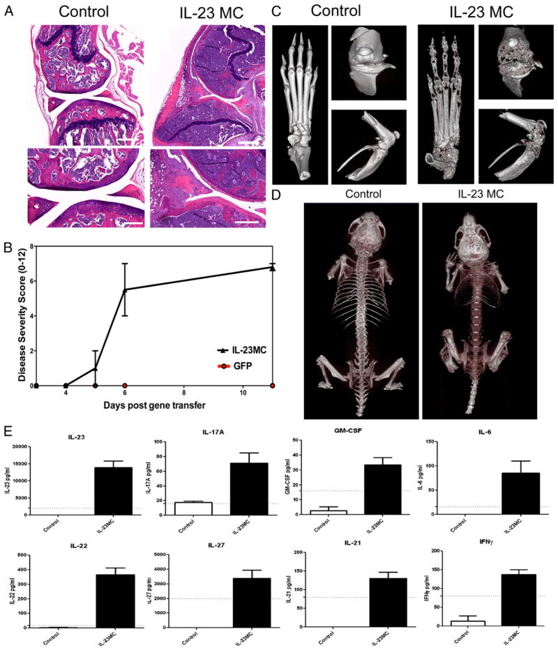
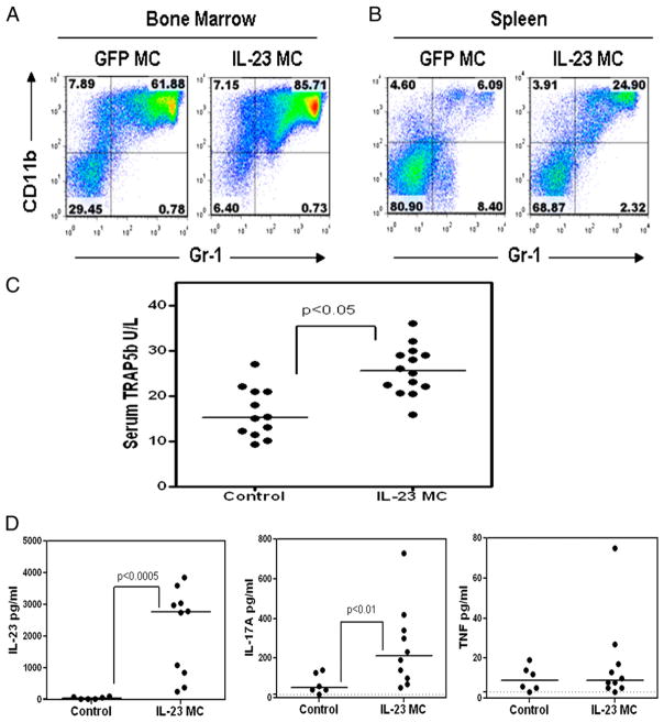
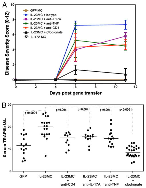
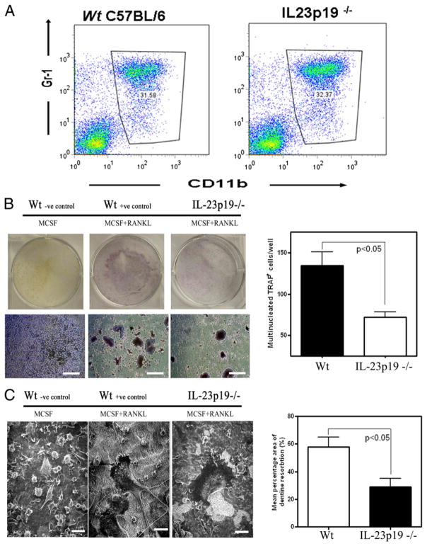
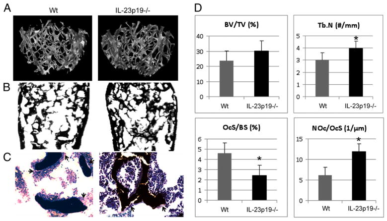
The role of IL-23 in the development of arthritis and bone metabolism was studied using systemic IL-23 exposure in adult mice via hydrodynamic delivery of IL-23 minicircle DNA in vivo and in mice genetically deficient in IL-23. Systemic IL-23 exposure induced chronic arthritis, severe bone loss, and myelopoiesis in the bone marrow and spleen, which resulted in increased osteoclast differentiation and systemic bone loss. The effect of IL-23 was partly dependent on CD4(+) T cells, IL-17A, and TNF, but could not be reproduced by overexpression of IL-17A in vivo. A key role in the IL-23-induced arthritis was made by the expansion and activity of myeloid cells. Bone marrow macrophages derived from IL-23p19(-/-) mice showed a slower maturation into osteoclasts with reduced tartrate-resistant acid phosphatase-positive cells and dentine resorption capacity in in vitro osteoclastogenesis assays. This correlated with fewer multinucleated osteoclast-like cells and more trabecular bone volume and number in 26-wk-old male IL-23p19(-/-) mice compared with control animals. Collectively, our data suggest that systemic IL-23 exposure induces the expansion of a myeloid lineage osteoclast precursor, and targeting IL-23 pathway may combat inflammation-driven bone destruction as observed in rheumatoid arthritis and other autoimmune arthritides.


Figures

FIGURE 1
FIGURE 1
Systemic IL-23 induces joint inflammation and extensive bone loss in vivo. A, H&E-stained decalcified sections of the knee joint showing extensive subchondral, metaphyseal, and epiphyseal bone loss, hyperplastic bone marrow, and pannus formation 30 d after IL-23 MC injection compared with GFP MC control. Scale bars, 500 μm (top panels) and 250 μm (bottom panels). Representative images of three independent experiments. B, Clinical disease severity score after IL-23 MC injection (average of four independent experiments). C, Three-dimensional rendered images from the paw and the anterior and side knee views 60 d after IL-23 MC injection (representative images from three independent experiments). D, Day 90 whole-body scan of control mice compared with the IL-23 MC-injected mice showing severe loss of bone mineral density in IL-23 MC-injected mice. E, Serum cytokine levels 28 d after IL-23 MC injection compared with GFP MC control mice. Cytokines presented were statistically different from control using serum data pooled from three independent experiments. Dotted line shows sensitivity of the assay. p < 0.05, Mann–Whitney nonparametric test.
FIGURE 2
FIGURE 2
Systemic IL-23 induces myelopoiesis, serum TRAP5b, and IL-17A. Flow cytometry of (A) bone marrow and (B) spleen 11 d after IL-23 MC injection compared with control GFP MC mice. C, Serum TRAP5b 11 d after IL-23 MC injection compared with GFP MC control (p < 0.05, Mann–Whitney nonparametric test; data pooled from four experiments). D, Serum IL-23, IL-17A, and TNF levels 11 d after IL-23 MC injection compared with GFP MC control (p < 0.05, Mann–Whitney nonparametric test; data pooled from three experiments).
FIGURE 3
FIGURE 3
Systemic IL-23–induced arthritis and elevated serum TRAP5b is inhibited by depletion of phagocytic cells. A, IL-23 MC induced arthritis after depletion of CD4 T cells or phagocytic cells, and neutralization of IL-17A or TNF. GFP MC and IL-17A MC controls also are presented (data pooled from three experiments). B, Serum TRAP5b 11 d after IL-23 MC injection (ANOVA).
FIGURE 4
FIGURE 4
IL-23p19−/− osteoclasts have impaired differentiation and function. A, Flow cytometry of bone marrow isolated from IL-23p19−/− or control mice cultured for 24 h in the presence of M-CSF. B, BMM isolated from IL-23p19−/− and control mice cultured for 6 d in the presence of M-CSF+RANKL form TRAP+ cells, which are also multinucleated (TRAP stain). Scale bars, 200 μm. C, Scanning electron photomicrographs of BMM cultures showing mature osteoclast resorbing activity (i.e., resorbed dentine has a rough, lighter color appearance). Scale bars, 50 μm. Representative data of three experiments done in triplicate.
FIGURE 5
FIGURE 5
Twenty-six–wk-old male IL-23–deficient mice have bone abnormalities caused by osteoclast dysregulation. A, Representative high-resolution μCT analysis of 26-wk-old male mouse’s trabecular bone in the distal femur. B, Von Kossa-stained sections of 26-wk-old male mouse’s distal femurs. Original magnification ×2. C, Toluidine blue counterstained von Kossa-stained sections of 26-wk-old male mouse’s distal femurs. Original magnification ×20. Black arrows indicate osteoclasts. D, BV/TV, Tb.N, OcS/BS, and OcN/OcS (NOc/OcS) measured in the distal femur. *p < 0.05.

References

    1. Teitelbaum SL. Bone resorption by osteoclasts. Science. 2000;289:1504–1508. - PubMed
    1. Lacey DL, Timms E, Tan HL, Kelley MJ, Dunstan CR, Burgess T, Elliott R, Colombero A, Elliott G, Scully S, et al. Osteoprotegerin ligand is a cytokine that regulates osteoclast differentiation and activation. Cell. 1998;93:165–176. - PubMed
    1. Simonet WS, Lacey DL, Dunstan CR, Kelley M, Chang MS, Lüthy R, Nguyen HQ, Wooden S, Bennett L, Boone T, et al. Osteoprotegerin: a novel secreted protein involved in the regulation of bone density. Cell. 1997;89:309–319. - PubMed
    1. Li J, Sarosi I, Yan X-Q, Morony S, Capparelli C, Tan H-L, McCabe S, Elliott R, Scully S, Van G, et al. RANK is the intrinsic hematopoietic cell surface receptor that controls osteoclastogenesis and regulation of bone mass and calcium metabolism. Proc Natl Acad Sci USA. 2000;97:1566–1571. - PMC - PubMed
    1. Kong YY, Feige U, Sarosi I, Bolon B, Tafuri A, Morony S, Capparelli C, Li J, Elliott R, McCabe S, et al. Activated T cells regulate bone loss and joint destruction in adjuvant arthritis through osteoprotegerin ligand. Nature. 1999;402:304–309. - PubMed

Publication types

MeSH terms

Substances