. 2011 Jul;121(7):2808-20.
doi: 10.1172/JCI44489. Epub 2011 Jun 13.

Engraftment of human nasal olfactory stem cells restores neuroplasticity in mice with hippocampal lesions

Affiliations

Engraftment of human nasal olfactory stem cells restores neuroplasticity in mice with hippocampal lesions

Emmanuel Nivet et al. J Clin Invest. 2011 Jul.

Abstract

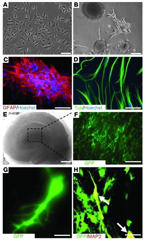
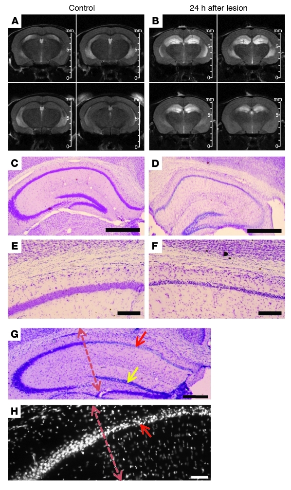
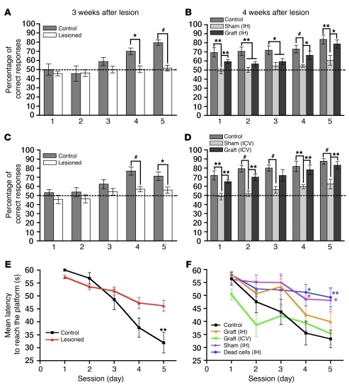
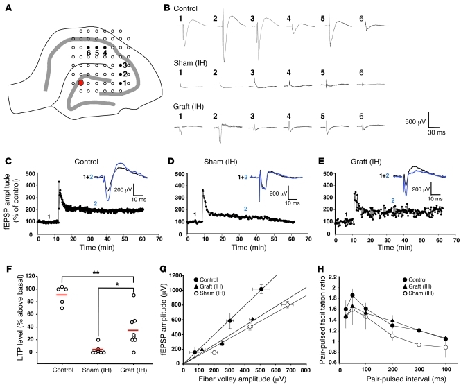
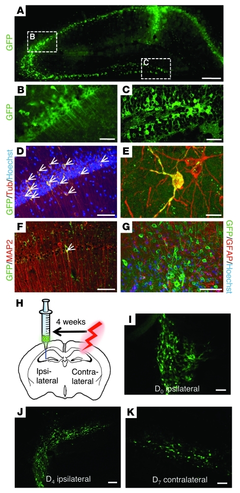
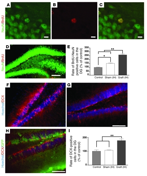
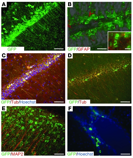
Stem cell-based therapy has been proposed as a potential means of treatment for a variety of brain disorders. Because ethical and technical issues have so far limited the clinical translation of research using embryonic/fetal cells and neural tissue, respectively, the search for alternative sources of therapeutic stem cells remains ongoing. Here, we report that upon transplantation into mice with chemically induced hippocampal lesions, human olfactory ecto-mesenchymal stem cells (OE-MSCs) - adult stem cells from human nasal olfactory lamina propria - migrated toward the sites of neural damage, where they differentiated into neurons. Additionally, transplanted OE-MSCs stimulated endogenous neurogenesis, restored synaptic transmission, and enhanced long-term potentiation. Mice that received transplanted OE-MSCs exhibited restoration of learning and memory on behavioral tests compared with lesioned, nontransplanted control mice. Similar results were obtained when OE-MSCs were injected into the cerebrospinal fluid. These data show that OE-MSCs can induce neurogenesis and contribute to restoration of hippocampal neuronal networks via trophic actions. They provide evidence that human olfactory tissue is a conceivable source of nervous system replacement cells. This stem cell subtype may be useful for a broad range of stem cell-related studies.


Figures

Figure 1
Figure 1. In vitro assessment of neurogenic characteristics of OE-MSCs.
Olfactory stem cells (A) gave rise to spherical clusters (B), expressing the GFAP neural stem cell marker (C), when grown in appropriate medium. Cells from spheres were then allowed to differentiate into neurons expressing the III–β-tubulin neuron marker (D). Freshly prepared mouse hippocampal slices were cultivated on culture insert (E) and loaded with sphere-derived GFP+ OE-MSCs (F). 3 weeks after culture at the air-liquid interface, some GFP+ OE-MSCs (green) adopted a neuron-like shape (G) and gave rise to MAP2-expressing neurons (H, white arrows). Scale bars: 100 μm (A, B, C, and D); 500 μm (E); 200 μm (F); 20 μm (G); 50 μm (H).
Figure 2
Figure 2. Lesion assessment.
24 hours after IH ibotenic acid injections, lesion extent was assessed in vivo by MRI (A and B). Examples of 4 axial contiguous T2-weighted images (slice thickness = 500 μm, TEeff = 60 ms, TR = 3000 ms, rare factor = 8; 8 averages) from control (A) and lesioned (B) mice are shown. In (B), the hypersignal (bright intensity) revealed the extent of the injury. Extent of ibotenic acid–induced neuronal death was visualized using cresyl violet staining (CF). When compared with controls (C and E), lesioned mice exhibited a dramatic cell loss in the whole hippocampus (D). High magnification images of CA1 pyramidal cell body layers in control and lesioned mouse are shown in E and F, respectively. In G (crystal violet staining) and H (Hoechst blue staining), region-specific lesions in stratum pyramidale of the CA1 (red arrow) and upper part of the stratum granulosum layer DG (yellow arrow) demonstrate their efficiency and specificity when compared with neighboring intact layers. Scale bars: 500 μm (C and D); 100 μm (E and F); 250 μm (G and H).
Figure 3
Figure 3. IH and ICV transplantation of human OE-MSCs improved hippocampus-dependent learning and memory.
Cognitive capacities of mice were assessed in the olfactory tubing maze (AD) and the Morris water maze (E and F). Associative or spatial memory in mice was assessed 3 weeks after lesion (A, C, and E) and 4 weeks after cell or culture medium transplantation (B, D, and F). (AD) Mean percentage of correct responses was obtained during 5 training sessions of 20 trials per day. (A and C) Lesioned mice (n = 2 × 16) exhibited significant impairment in an associative memory task when compared with control mice (n = 2 × 8). 4 weeks after cell implantation in the lesioned sites (B) or in the lateral ventricles (D), grafted mice (grafted, n = 2 × 8) demonstrated a significant improvement in associative memory when compared with vehicle-grafted mice (sham-grafted, n = 2 × 8). (E and F) Graphs showing the mean latencies to reach the platform during 5 training sessions of 4 trials per day. (E) Lesioned mice (n = 32) exhibited significant impairment in a visuospatial learning task when compared with control mice (n = 8). 4 weeks after cell implantation in the lesioned sites (grafted IH, n = 8) or in the lateral ventricles (grafted ICV, n = 8), grafted mice demonstrated a significant improvement in spatial learning and memory when compared with vehicle-grafted mice (sham-grafted and dead cells, n = 8, respectively). See also Supplemental Table 1 and Supplemental Videos 1 and 2. *P < 0.05; **P < 0.01; #P < 0.001.
Figure 4
Figure 4. Recovery of excitatory synaptic transmission and LTP after human OE-MSC transplantation in lesioned hippocampi.
5 weeks after cell grafting, acute hippocampal slices were prepared and synaptic transmission was evaluated with a multi-electrode array. (A) Schematic diagram illustrating the positioning of hippocampal slices on a 60-electrode array. fEPSPs were evoked along the hippocampal circuitry by delivering stimulations (red electrode) in the DG. Recording electrodes (1 to 6) were located in CA1 and CA3 subfields. (B) Representative fEPSPs from control, sham-grafted, and grafted mice. A partial recovery of evoked fEPSPs was observed in slices from grafted but not in vehicle-treated mice. (CF) The same setting was used to record CA1 LTP. Illustrative examples of LTP triggered by high-frequency stimulation are shown in control (C), sham-grafted (D), and grafted (E) mice. fEPSP amplitude was normalized against control values and plotted. (F) Group data for LTP in control (n = 5), sham-grafted (n = 7), and grafted (n = 8) mice; mean values of LTP levels (measured 40 minutes after HFS) are indicated with an horizontal bar (each animal is represented by a circle). (G) Basal synaptic transmission in control (n = 4), sham-grafted (n = 4), and grafted (n = 4) mice. The input/output curve was generated by applying increasing stimulation intensities and by plotting the fEPSP amplitude as a function of the corresponding fiber volley amplitude. (H) Group data for paired-pulse facilitation in control (n = 4), sham-grafted (n = 4), and grafted (n = 4) mice. *P < 0.05; **P < 0.01.
Figure 5
Figure 5. Human OE-MSCs transplanted into lesioned mouse hippocampi survived, migrated, and differentiated into neurons.
(A) 5 weeks after transplantation, exogenous GFP+ human OE-MSCs were present in the different fields (CA1, CA3, DG) of the lesioned hippocampus. (B and C) GFP+ human OE-MSCs were mostly found in pyramidal (CA3, B) and granule cell layers (DG, C). (D) Within these layers, a high proportion (69%) of GFP+ human OE-MSCs (green) expressed III–β-tubulin (red) (white arrows in D). (E) High magnification of the merged picture of human GFP+ OE-MSCs (green) expressing III–β-tubulin (red). (F) A small proportion of human GFP+ OE-MSCs (green) expressed MAP2 (white arrow), a marker for mature neurons (red). (G) No GFP+ human OE-MSC (green) was ever found to express the astrocytic marker GFAP (red). (H) 4 weeks after lesioning the right hippocampus, GFP+ OE-MSCs were transplanted into the intact hippocampus (i.e., left hemisphere). At day 0 (D0) after transplantation, cells formed clusters and were only observed within the injection site as a cell cluster (I). At D4 after transplantation, numerous GFP+ cells were observed migrating outside the injection site toward the contralateral lesioned hippocampus (J). At D7 after transplantation, few GFP+ OE-MSCs were observed inside the contralateral hippocampus (K). Scale bars: 250 μm (A); 200 μm (J); 100 μm (B, C, D, F, G, I, and K); 20 μm (E). See also Supplemental Video 3.
Figure 6
Figure 6. Human OE-MSCs stimulated endogenous neurogenesis after transplantation in lesioned hippocampi.
5 weeks after grafting, brain sections of mice injected with BrdU twice a day during 3 days following cell implantation (n = 5 for each group) were immunostained with anti-NeuN (green) and anti-BrdU antibodies (red) (AD). Quantification of BrdU+/NeuN+ cells in DG indicated an increased number of mitotic cells in grafted (IH) mice when compared with sham-grafted (IH) (P < 0.05) and control (P < 0.01) mice (E). DCX immunohistochemistry revealed the presence of immature neurons in the DG of control (G) and grafted (IH) (F, H) mice. As shown in (H), no GFP+ human OE-MSC was found to express DCX. Lesioned mice with IH transplant of human OE-MSCs (grafted [IH], n = 5) exhibited a higher percentage of DCX-positive cells when compared with vehicle-treated (sham-grafted [IH], n = 5) and control (n = 5) mice (I). (E and I). Scale bars: 10 μm (AC); 100 μm (D, F, G, and H). *P < 0.05; **P < 0.01.
Figure 7
Figure 7. Human OE-MSCs transplanted into the cerebrospinal fluid (CSF) of hippocampus-lesioned mice survived at least 5 weeks, migrated, and differentiated into neurons.
(A) Increased density of GFP+ cells within the pyramidal cell body layers demonstrated the ability of human OE-MSCs to migrate from the CSF toward the injury zone. (B) As demonstrated by confocal image reconstitution using projection transparency (see also Supplemental Video 4), exogenous GFP+ cells were also remarkably distributed within the granule cell body layers of the DG. The insert indicates that not a single human OE-MSC expressed the astrocytic marker GFAP (red). (C and D) Within the CA3 field, some pyramidal cell–like and interneuron-like GFP+ human OE-MSCs (green) expressed III–β-tubulin (red) (white arrows in C, yellow arrow in D), but others were immunonegative for this immature neuronal marker (green arrows in D). (E) Exogenous GFP+ cells migrated as well in other cerebral areas (cortical area in E), but remained immunonegative for the mature neuronal marker MAP2 (red). (F) GFP+ cells exhibiting an undifferentiated morphology were found at the margin of the ventricular areas. Scale bars: 100 μm (A, C, D, E, and F); 30 μm (B, insert). See also Supplemental Video 4.

Similar articles

Cited by

References

    1. Maguire EA, et al. Navigation-related structural change in the hippocampi of taxi drivers. Proc Natl Acad Sci U S A. 2000;97(8):4398–4403. doi: 10.1073/pnas.070039597. - DOI - PMC - PubMed
    1. Wiltrout C, Lang B, Yan Y, Dempsey RJ, Vemuganti R. Repairing brain after stroke: a review on post–ischemic neurogenesis. Neurochem Int. 2007;50(7–8):1028–1041. - PubMed
    1. Jin K, et al. Increased hippocampal neurogenesis in Alzheimer’s disease. Proc Natl Acad Sci U S A. 2004;101(1):343–347. doi: 10.1073/pnas.2634794100. - DOI - PMC - PubMed
    1. Li B, et al. Failure of neuronal maturation in Alzheimer disease dentate gyrus. J Neuropathol Exp Neurol. 2008;67(1):78–84. doi: 10.1097/nen.0b013e318160c5db. - DOI - PMC - PubMed
    1. Lindvall O, Kokaia Z. Stem cells for the treatment of neurological disorders. Nature. 2006;441(7097):1094–1096. - PubMed

Publication types

MeSH terms