. 2011 Jul;121(7):2736-49.
doi: 10.1172/JCI45444. Epub 2011 Jun 13.

Krüppel-like factor 4 regulates macrophage polarization

Affiliations

Krüppel-like factor 4 regulates macrophage polarization

Xudong Liao et al. J Clin Invest. 2011 Jul.

Abstract

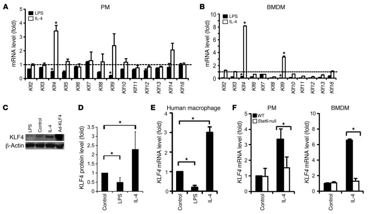
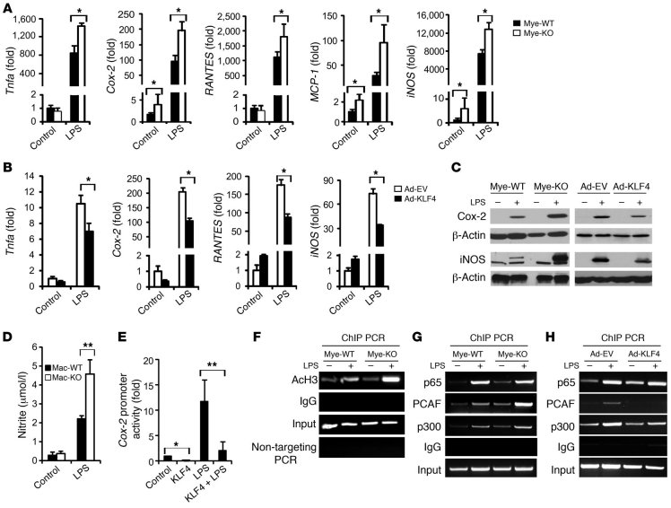
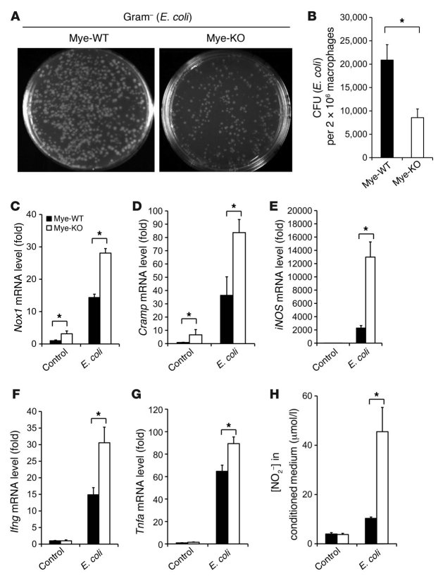
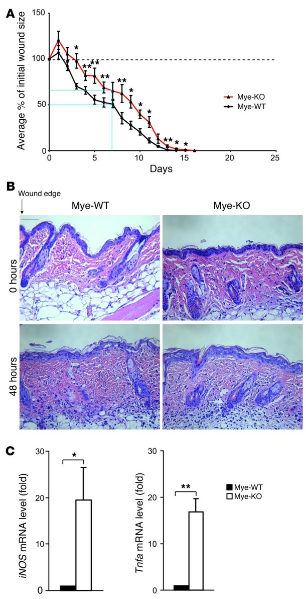
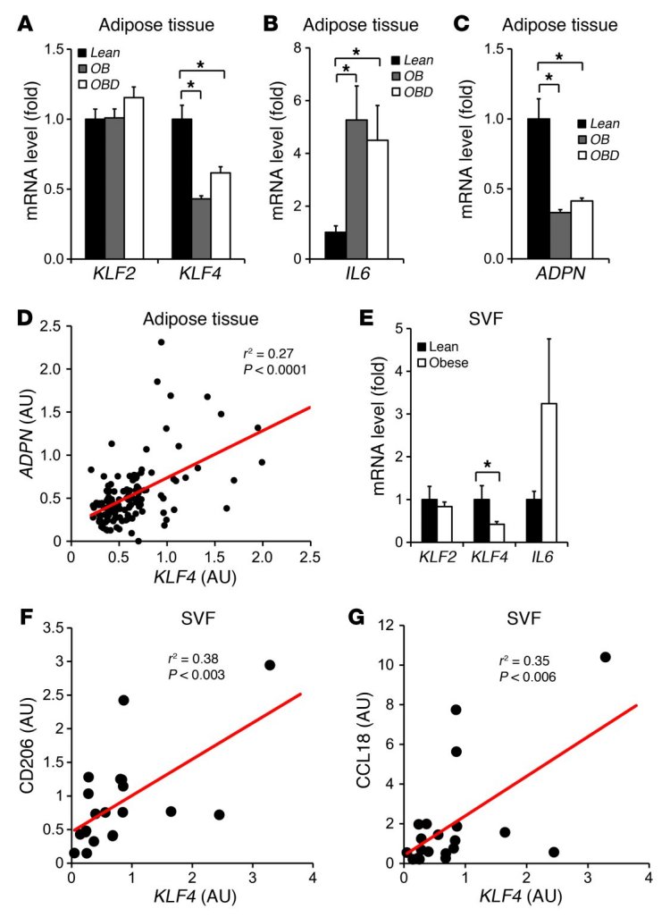
Current paradigms suggest that two macrophage subsets, termed M1 and M2, are involved in inflammation and host defense. While the distinct functions of M1 and M2 macrophages have been intensively studied - the former are considered proinflammatory and the latter antiinflammatory - the determinants of their speciation are incompletely understood. Here we report our studies that identify Krüppel-like factor 4 (KLF4) as a critical regulator of macrophage polarization. Macrophage KLF4 expression was robustly induced in M2 macrophages and strongly reduced in M1 macrophages, observations that were recapitulated in human inflammatory paradigms in vivo. Mechanistically, KLF4 was found to cooperate with Stat6 to induce an M2 genetic program and inhibit M1 targets via sequestration of coactivators required for NF-κB activation. KLF4-deficient macrophages demonstrated increased proinflammatory gene expression, enhanced bactericidal activity, and altered metabolism. Furthermore, mice bearing myeloid-specific deletion of KLF4 exhibited delayed wound healing and were predisposed to developing diet-induced obesity, glucose intolerance, and insulin resistance. Collectively, these data identify KLF4 as what we believe to be a novel regulator of macrophage polarization.


Figures

Figure 1
Figure 1. Klf4 expression is augmented by M2 and inhibited by M1 stimuli in macrophages.
(A and B) KLF mRNA levels in (A) mouse PMs and (B) BMDMs after 16 hours of stimulation with LPS (50 ng/ml) or mouse IL-4 (5 ng/ml). Gene expression levels were assessed by qPCR and normalized to those in untreated cells (dashed line). Klf1, Klf15, and Klf17 were not detectable in PMs or BMDMs. Klf5 and Klf14 were not detectable in BMDMs. n = 5. (C and D) KLF4 protein levels after LPS and IL-4 treatment. n = 3. (E) KLF4 mRNA level in M-CSF–differentiated primary human macrophages (from 3 donors) stimulated with LPS (50 ng/ml) or human IL-4 (10 ng/ml) for 16 hours. (F) IL-4–mediated induction of the Klf4 gene was diminished in Stat6-null PMs and BMDMs. n = 3. C57BL/6J mice were used as control (WT). *P < 0.05, Student’s t test.
Figure 2
Figure 2. KLF4 is essential for IL-4–mediated macrophage M2 polarization.
(A) Impairment of M2 marker gene expression in KLF4-deficient macrophages. PMs isolated from Mye-WT and Mye-KO mice were treated with IL-4 for 16 hours. n = 3 in each group. (B) KLF4 overexpression enhances M2 gene expression. RAW264.7 cells were infected with either Ad-GFP (Ad-EV) or Ad-KLF4 for 24 hours prior to treatment with IL-4 for 16 hours. n = 3 in each group. (C) Representative Western blot showing IL-4–mediated protein induction of Arg-1, Retnla, Chi3l3, and PPARγ in Mye-WT and Mye-KO macrophages. (D) Quantification of Western blot data by densitometry. For Arg-1, protein levels were normalized to Mye-WT control group. For Retnla, Chi3l3 and PPARγ, only IL-4–induced protein levels were calculated and normalized to the IL-4–treated Mye-WT group, due to extremely low levels of expression at baseline. Data were calculated from 3–5 independent blots. (E) Synergistic activation of the mouse Arg1 promoter by KLF4 and Stat6 as assayed by transient transfection. WT, ~4 kb WT mouse Arg1 promoter; ΔKLF, Arg1 promoter with both KLF-binding sites mutated; ΔStat6: Arg1 promoter with Stat6-binding site mutated. Transient transfection experiments were performed in RAW264.7 cells. n = 3. (F) KLF4 binding to Arg1 promoter detected by ChIP assay in WT and Stat6-null BMDMs with or without IL-4 treatment (4 hours). *P < 0.05, **P < 0.01, Student’s t test with Bonferroni correction.
Figure 3
Figure 3. KLF4 deficiency enhances macrophage M1 polarization.
(A) Enhanced expression of M1 marker genes in KLF4-deficient macrophages stimulated by LPS for 16 hours. n = 3 in each group. (B) Attenuated expression of M1 marker genes in RAW264.7 cells with overexpression of KLF4. n = 3 in each group. (C) Protein levels of Cox-2 and iNOS in PMs (left) and RAW264.7 cells (right). (D) KLF4-deficient macrophages generate more NO after LPS stimulation. n = 3 in each group. (E) Inhibition of Cox-2 promoter by KLF4. Transient transfection experiments were performed in RAW264.7 cells. n = 3. (F) Enhanced acetylation of histone H3 (AcH3) in LPS-treated KLF4-deficient macrophages demonstrated by ChIP. (G and H) KLF4 regulates LPS-induced recruitment of p300/PCAF as shown by deficiency (G) and overexpression (H) experiments. *P < 0.05, **P < 0.01, Student’s t test with Bonferroni correction.
Figure 4
Figure 4. KLF4-deficient macrophages exhibit enhanced bactericidal activity.
(A and B) KLF4-deficient macrophages exhibit enhanced bactericidal activity against E. coli ex vivo. n = 3 in each group. (CG) Expression of genes involved in bactericidal activity is augmented in KLF4-deficient macrophages after incubation with E. coli. n = 3 in each group. (H) NO production in macrophage was determined by nitrite (NO2) levels in conditioned medium. n = 3 in each group. *P < 0.05, Student’s t test with Bonferroni correction.
Figure 5
Figure 5. Myeloid KLF4 deficiency results in delayed wound healing.
(A) Wound healing graphs of the mean percentage of initial wound area demonstrate a delay in wound healing in Mye-KO mice. At 1 week after wounding, Mye-WT mice had a 49.3% total reduction in average wound size, in contrast to a 34.9% reduction in Mye-KO mice (blue lines). Furthermore, complete wound closure in Mye-KO mice occurred on day 16, compared with day 14 for WT mice. Dashed line at 100% indicates “baseline” percentage of wound area. n = 9 in each group. (B) H&E staining of skin surround wound edge at 48 hours demonstrates similar levels of myeloid cells in the perilesional wound tissue. Scale bar: 150 μm. (C) Expression of iNOS and Tnfa from skin surrounding the wound edge as assessed by qPCR at 48 hours after wounding. n = 3 per genotype. *P < 0.05, **P < 0.01, Student’s t test.
Figure 6
Figure 6. Relationship of KLF4 to obesity in human subjects.
(AC) Gene expression of KLF2, KLF4, IL6, and ADPN in subcutaneous adipose tissue (needle biopsies) from 48 nondiabetic obese (OB), 37 obese diabetic (OBD), and 20 control (Lean) subjects. (D) Significant correlation between gene expression of KLF4 and ADPN in human adipose tissue. r2 and P values by Pearson correlation test. (E) KLF2, KLF4, and IL6 gene expression in SVF from 9 non-obese and 12 obese patients. (F and G) Significant correlations between gene expression of KLF4 and CD206 and CCL18 in SVF from 9 non-obese and 12 obese patients. r2 and P values by Pearson correlation test. *P < 0.05, Kruskal-Wallis test (vs. lean group). Gene expression in SVF samples was normalized to CD68.
Figure 7
Figure 7. Myeloid-specific deficiency of KLF4 exaggerates HFD-induced obesity and insulin resistance state in mice.
(A) Impairment of IL-4–mediated lipid uptake (left panel) and β-oxidation (right panel) in KLF4-deficient macrophages. [3H] counts were normalized to protein content and expressed as fold increase relative to untreated controls. n = 8 in each group. (B) KLF4-deficient macrophages exhibit enhanced glucose uptake before and after LPS stimulation (left panel) and release more L(+)-lactate (right panel), a product of glycolysis. [3H] counts from 2-deoxy-[3H]-d-glucose in the cell lysate and L(+)-lactate concentration in the conditioned medium were normalized to protein content and expressed as fold increase over untreated controls. n = 8 in each group. (C and D) Mye-KO mice on HFD gained more weight (C) and stored more fat (D) than Mye-WT controls. n = 6 in each group. Subc., subcutaneous. (E and F) After 10–12 weeks of HFD, Mye-KO mice show impaired glucose metabolism as revealed by glucose intolerance (E) and resistance to exogenous insulin (F). n = 6 per genotype. *P < 0.05, **P < 0.01, Student’s t test with Bonferroni correction.
Figure 8
Figure 8. Mye-KO mice exhibit altered inflammatory and metabolic gene expression in tissues following HFD.
(A and B) Myeloid deficiency of KLF4 in HFD-mice affects transcription of many genes involved in nutrition uptake, β-oxidation, oxidative phosphorylation and inflammation in white adipose tissue (A) and skeletal muscle (B). n = 6 in each group. (C) Increased macrophage infiltration of white fat tissues and muscle tissues as determined by Mac-3 antibody staining. Original magnification, ×400. (D) Quantification of Mac-3 staining. n = 6 in each group. (E) Myeloid deficiency of KLF4 in HFD-fed mice affects expression of macrophage M1 and M2 genes in SVF. n = 6 in each group. *P < 0.05, Student’s t test. (F) Schematic of proposed mechanism. See text for details.

References

    1. Heilbronn LK, Campbell LV. Adipose tissue macrophages, low grade inflammation and insulin resistance in human obesity. Curr Pharm Des. 2008;14(12):1225–1230. - PubMed
    1. Johnson JL, Newby AC. Macrophage heterogeneity in atherosclerotic plaques. Curr Opin Lipidol. 2009;20(5):370–378. - PMC - PubMed
    1. Mantovani A, Sozzani S, Locati M, Allavena P, Sica A. Macrophage polarization: tumor-associated macrophages as a paradigm for polarized M2 mononuclear phagocytes. Trends Immunol. 2002;23(11):549–555. doi: 10.1016/S1471-4906(02)02302-5. - DOI - PubMed
    1. Charo IF. Macrophage polarization and insulin resistance: PPARgamma in control. Cell Metab. 2007;6(2):96–98. - PubMed
    1. Evans TJ. The role of macrophages in septic shock. Immunobiology. 1996;195(4–5):655–659. - PubMed

Publication types

MeSH terms